Department of Cardiology, School of Cardiovascular Medicine and Sciences, British Heart Foundation Centre for Research Excellence, Faculty of Life Sciences and Medicine, The Rayne Institute, King's College London, St Thomas's Hospital, Lambeth Palace Road, London SE1 7EH, UK.
Randall Division of Cell and Molecular Biophysics, King's College London, New Hunt's House, Guy's Hospital Campus, London SE1 1UL, UK.
Cardiovasc Res. 2018 Jan 1;114(1):138-157. doi: 10.1093/cvr/cvx206.
PKN1 is a stress-responsive protein kinase acting downstream of small GTP-binding proteins of the Rho/Rac family. The aim was to determine its role in endogenous cardioprotection.
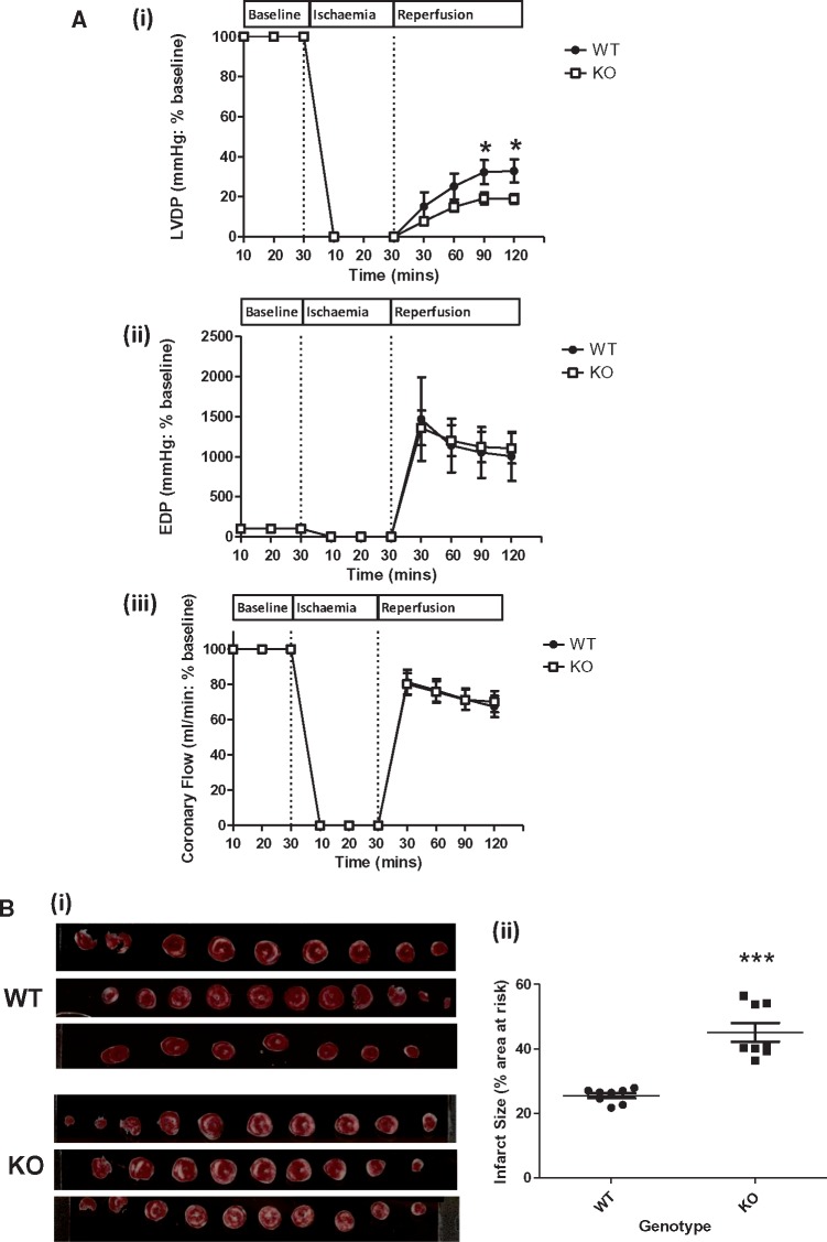
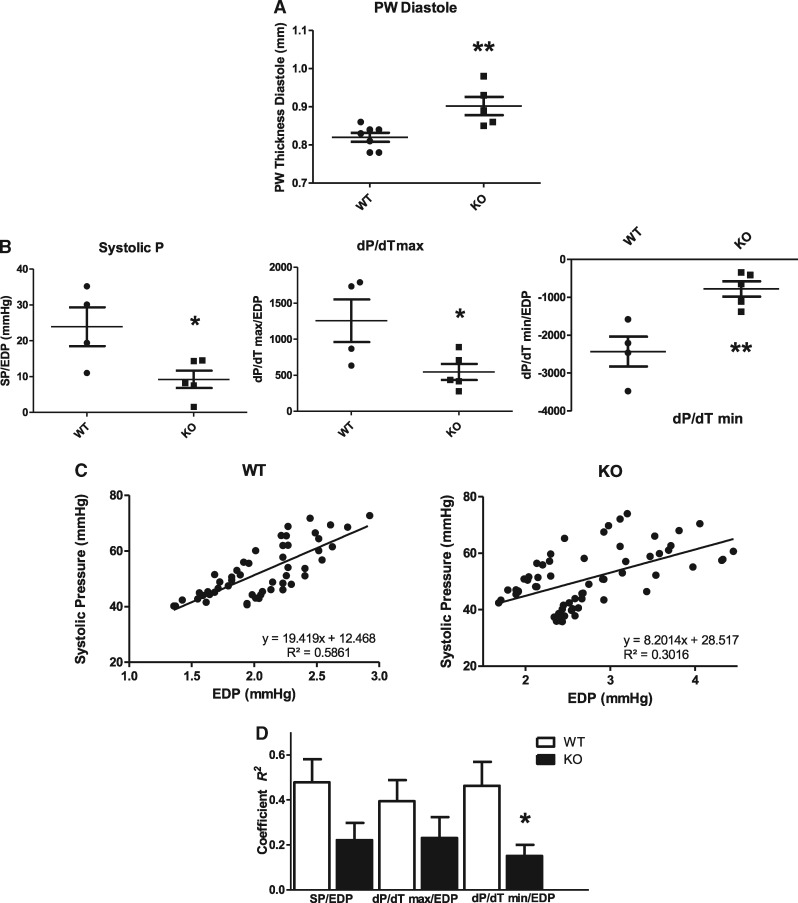
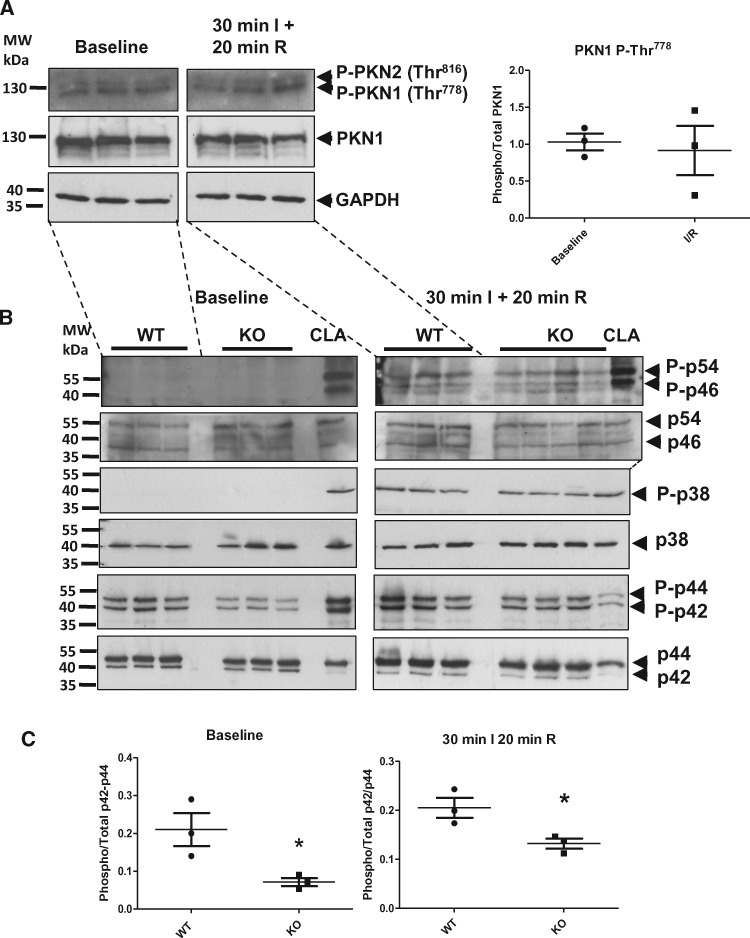
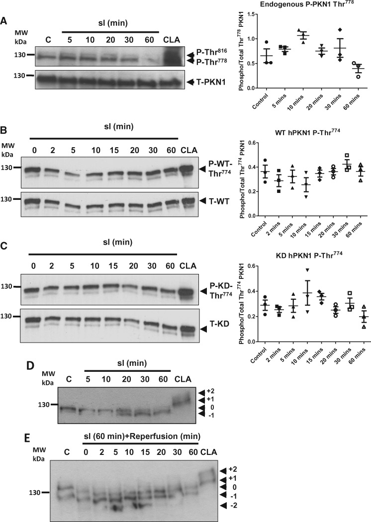
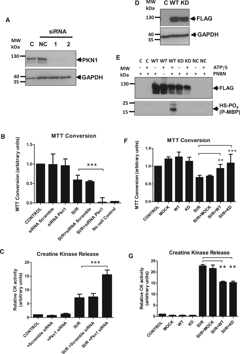
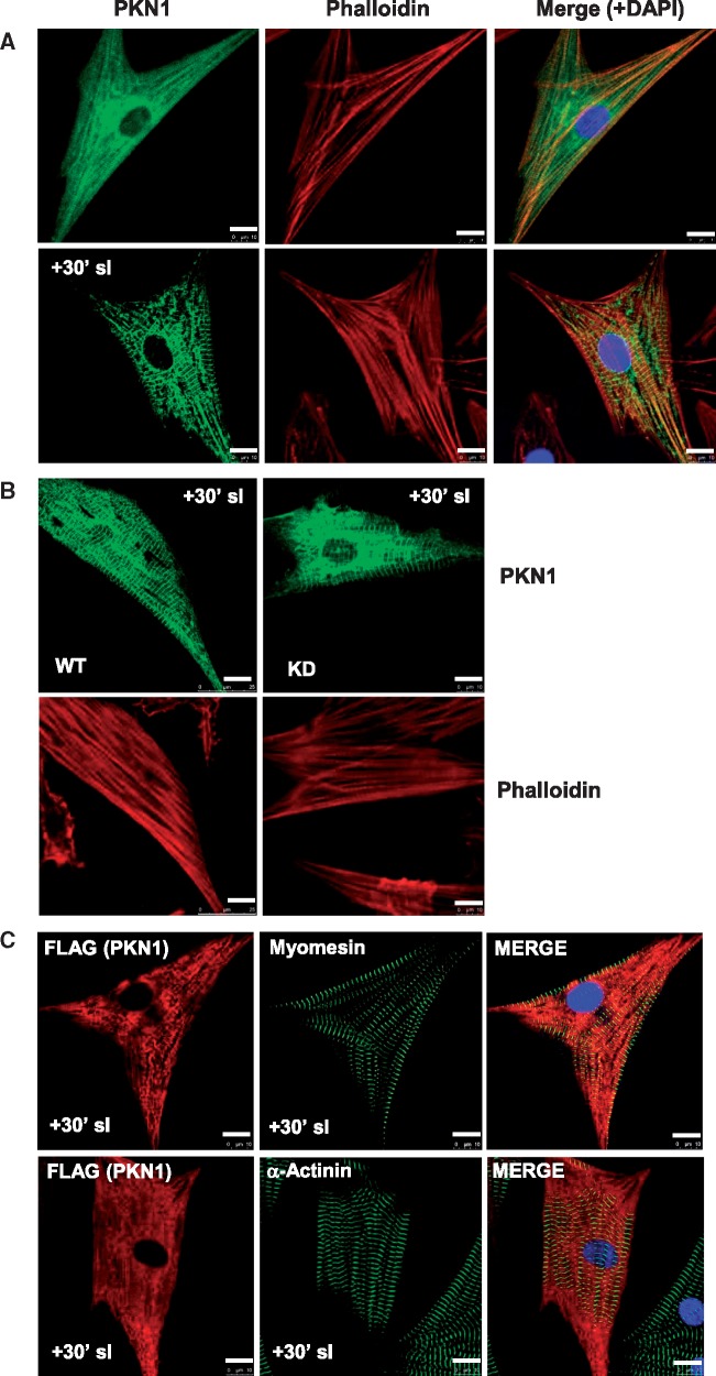
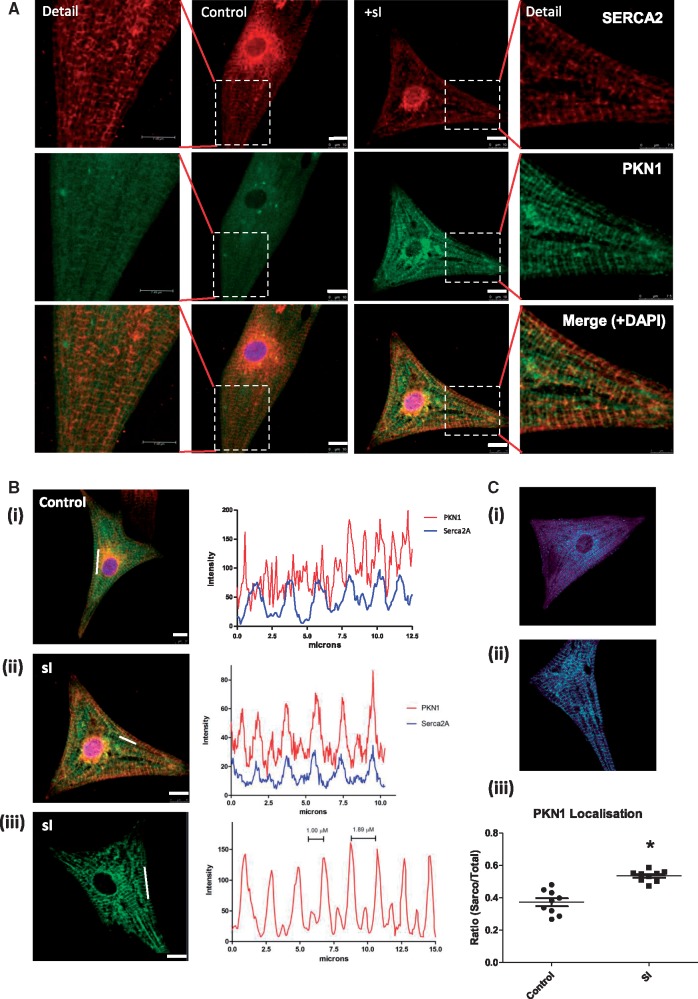
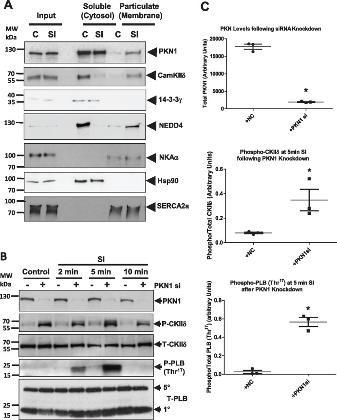
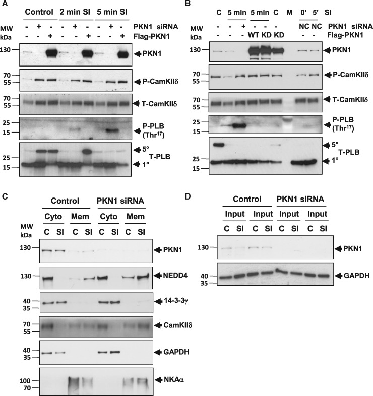
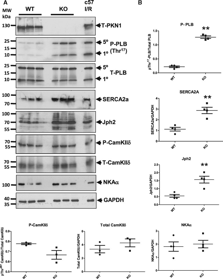
Hearts from PKN1 knockout (KO) or wild type (WT) littermate control mice were perfused in Langendorff mode and subjected to global ischaemia and reperfusion (I/R). Myocardial infarct size was doubled in PKN1 KO hearts compared to WT hearts. PKN1 was basally phosphorylated on the activation loop Thr778 PDK1 target site which was unchanged during I/R. However, phosphorylation of p42/p44-MAPK was decreased in KO hearts at baseline and during I/R. In cultured neonatal rat ventricular cardiomyocytes (NRVM) and NRVM transduced with kinase dead (KD) PKN1 K644R mutant subjected to simulated ischaemia/reperfusion (sI/R), PhosTag® gel analysis showed net dephosphorylation of PKN1 during sI and early R despite Thr778 phosphorylation. siRNA knockdown of PKN1 in NRVM significantly decreased cell survival and increased cell injury by sI/R which was reversed by WT- or KD-PKN1 expression. Confocal immunofluorescence analysis of PKN1 in NRVM showed increased localization to the sarcoplasmic reticulum (SR) during sI. GC-MS/MS and immunoblot analysis of PKN1 immunoprecipitates following sI/R confirmed interaction with CamKIIδ. Co-translocation of PKN1 and CamKIIδ to the SR/membrane fraction during sI correlated with phospholamban (PLB) Thr17 phosphorylation. siRNA knockdown of PKN1 in NRVM resulted in increased basal CamKIIδ activation and increased PLB Thr17 phosphorylation only during sI. In vivo PLB Thr17 phosphorylation, Sarco-Endoplasmic Reticulum Ca2+ ATPase (SERCA2) expression and Junctophilin-2 (Jph2) expression were also basally increased in PKN1 KO hearts. Furthermore, in vivo P-V loop analysis of the beat-to-beat relationship between rate of LV pressure development or relaxation and end diastolic P (EDP) showed mild but significant systolic and diastolic dysfunction with preserved ejection fraction in PKN1 KO hearts.
Loss of PKN1 in vivo significantly reduces endogenous cardioprotection and increases myocardial infarct size following I/R injury. Cardioprotection by PKN1 is associated with reduced CamKIIδ-dependent PLB Thr17 phosphorylation at the SR and therefore may stabilize the coupling of SR Ca2+ handling and contractile function, independent of its kinase activity.
PKN1 是一种应激反应蛋白激酶,作用于 Rho/Rac 家族小 GTP 结合蛋白的下游。本研究旨在确定其在内源性心脏保护中的作用。
PKN1 敲除(KO)或野生型(WT)同窝对照小鼠的心脏在 Langendorff 模式下进行灌注,并进行整体缺血再灌注(I/R)。与 WT 心脏相比,PKN1 KO 心脏的心肌梗死面积增加了一倍。PKN1 在激活环 Thr778 PDK1 靶位上基础磷酸化,I/R 过程中无变化。然而,KO 心脏在基础状态和 I/R 过程中 p42/p44-MAPK 的磷酸化减少。在培养的新生大鼠心室肌细胞(NRVM)和转导激酶失活(KD)PKN1 K644R 突变体的 NRVM 中,进行模拟缺血/再灌注(sI/R)时,PhosTag®凝胶分析显示尽管 Thr778 磷酸化,但在 sI 和早期 R 期间 PKN1 净去磷酸化。NRVM 中的 PKN1 siRNA 敲低显著降低了 sI/R 引起的细胞存活率并增加了细胞损伤,而 WT 或 KD-PKN1 的表达则逆转了这种损伤。NRVM 中 PKN1 的共聚焦免疫荧光分析显示,在 sI 期间 PKN1 定位于肌浆网(SR)。sI/R 后 PKN1 免疫沉淀的 GC-MS/MS 和免疫印迹分析证实与 CamKIIδ 相互作用。在 sI 期间,PKN1 和 CamKIIδ 共转位到 SR/膜部分与肌球蛋白轻链磷酸酶(PLB) Thr17 磷酸化相关。NRVM 中的 PKN1 siRNA 敲低导致基础状态下 CamKIIδ 激活增加,仅在 sI 期间 PLB Thr17 磷酸化增加。体内 PKN1 KO 心脏的 PLB Thr17 磷酸化、肌浆网-内质网 Ca2+ATP 酶(SERCA2)表达和连接蛋白-2(Jph2)表达也基础增加。此外,LV 压力发展或松弛与舒张末期 P(EDP)之间的逐个搏动关系的 P-V 环分析显示,PKN1 KO 心脏的收缩和舒张功能轻度但显著受损,射血分数保留。
体内 PKN1 的缺失显著降低了缺血再灌注损伤后的内源性心脏保护作用,并增加了心肌梗死面积。PKN1 的心脏保护作用与减少 CamKIIδ 依赖性 PLB Thr17 在 SR 上的磷酸化有关,因此可能稳定 SR Ca2+ 处理和收缩功能的偶联,而不依赖其激酶活性。