Department of Veterinary Pathology, Faculty of Veterinary Medicine, University of Zagreb, Vjekoslava Heinzela 55, 10000, Zagreb, Croatia.
Faculty of Veterinary Medicine, University of Zagreb, Vjekoslava Heinzela 55, 10000, Zagreb, Croatia.
Parasit Vectors. 2017 Oct 18;10(1):495. doi: 10.1186/s13071-017-2412-1.
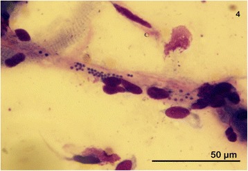
Classification of Babesia parasites has traditionally relied on morphological differentiation based on piroplasm size and shape. Molecular typing has subsequently revealed a more complex taxonomy for these piroplasms than previously thought. To evaluate the factors that influence the morphology of Babesia species upon microscopic examination and hence, their taxonomic classification, we performed detailed characterizations of piroplasms from archival and prospective collections of cytological samples of dogs with piroplasmosis before and after death. Merozoite morphology and time of parasite disappearance following imidocarb dipropionate was also investigated.
The study was divided into a (i) review of archived cytological slides from confirmed cases of canine piroplasmosis, and (ii) a prospective study of smears and tissue imprints from 15 recently necropsied dogs. The latter group could be further sub-divided into a non-treated group and an imidocarb dipropionate-treated group. Exact times of treatment before death were reviewed. Additional blood smears prepared from the live dogs and taken before therapy were also evaluated in the latter group. Parasite burden per each slide was determined in both studies. The shape and size of merozoites were described from blood smears taken while the dogs were alive and from different organs during necropsy. The results of all measurements were statistically analyzed.
The morphology and size of merozoites from live dogs corresponded to that of previously described 'large' Babesia. The morphology and size of merozoites were significantly different (P < 0.001) in postmortem samples, however, and more consistent in shape and size with piroplasm cells previously referred to as 'small' Babesia. PCR and sequencing confirmed B. canis as the causative agent of disease in all investigated dogs, including in postmortem negative tissue imprints from dogs treated at least 24 h before death.
Changes in the morphology of 'large' B. canis to 'small'-like Babesia observed by light microscopy appear to represent a common postmortem change. Classification of Babesia parasites into 'large' and 'small' Babesia using only microscopy of postmortem slides should be treated with caution. PCR-based methodologies for detection and molecular typing of Babesia spp. may prove valuable for investigating suspected cases of babesiosis following necropsy.
巴贝虫寄生虫的分类传统上依赖于基于血孢子大小和形状的形态学分化。随后的分子分型揭示了这些巴贝斯虫比以前认为的更为复杂的分类。为了评估影响巴贝斯虫物种在显微镜检查下形态的因素,从而影响其分类,我们对存档和前瞻性收集的死于巴贝斯虫病的犬的细胞学样本进行了详细的巴贝斯虫特征描述。还研究了抗焦磷酸二甲酯治疗前后裂殖体形态和寄生虫消失时间。
该研究分为(i)回顾确诊的犬巴贝斯虫病存档细胞学载玻片,和(ii)对 15 只最近尸检犬的涂片和组织印模进行前瞻性研究。后者可以进一步细分为未治疗组和抗焦磷酸二甲酯治疗组。回顾了死前治疗的确切时间。还评估了后者组中来自活犬的额外血涂片,并在治疗前进行了评估。在两项研究中,均确定了每个载玻片的寄生虫负担。在犬存活期间以及尸检时从不同器官采集血液涂片,描述裂殖体的形状和大小。对所有测量结果进行了统计学分析。
来自活犬的裂殖体的形态和大小与先前描述的“大”巴贝斯虫相符。然而,死后样本中的裂殖体形态和大小有显著差异(P < 0.001),与先前称为“小”巴贝斯虫的类血孢子细胞形态和大小更一致。PCR 和测序证实所有研究犬均为犬巴贝斯虫感染,包括至少在死前 24 小时接受治疗的死后组织印模阴性犬。
通过光镜观察到的“大”犬巴贝斯虫形态向“小”类巴贝斯虫的变化似乎代表了一种常见的死后变化。仅通过死后载玻片的显微镜检查将巴贝斯虫寄生虫分类为“大”和“小”巴贝斯虫应谨慎对待。PCR 检测和分子分型方法可能对死后怀疑巴贝斯虫病的调查有价值。