Herbst Elmar, Albers Marcio, Burnham Jeremy M, Fu Freddie H, Musahl Volker
Department of Orthopaedic Surgery, University of Pittsburgh Medical Center, Pittsburgh, Pennsylvania, USA.
Department of Orthopaedic Sports Medicine, Klinikum rechts der Isar, Technical University Munich, Munich, Germany.
Orthop J Sports Med. 2017 Oct 6;5(10):2325967117730805. doi: 10.1177/2325967117730805. eCollection 2017 Oct.
Significant controversy exists regarding the anterolateral structures of the knee.
To determine the layer-by-layer anatomic structure of the anterolateral complex of the knee.
Descriptive laboratory study.
Twenty fresh-frozen cadaveric knees (age range, 38-56 years) underwent a layer-by-layer dissection to systematically expose and identify the various structures of the anterolateral complex. Quantitative measurements were performed, and each layer was documented with high-resolution digital imaging.
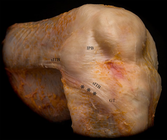
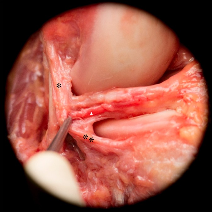
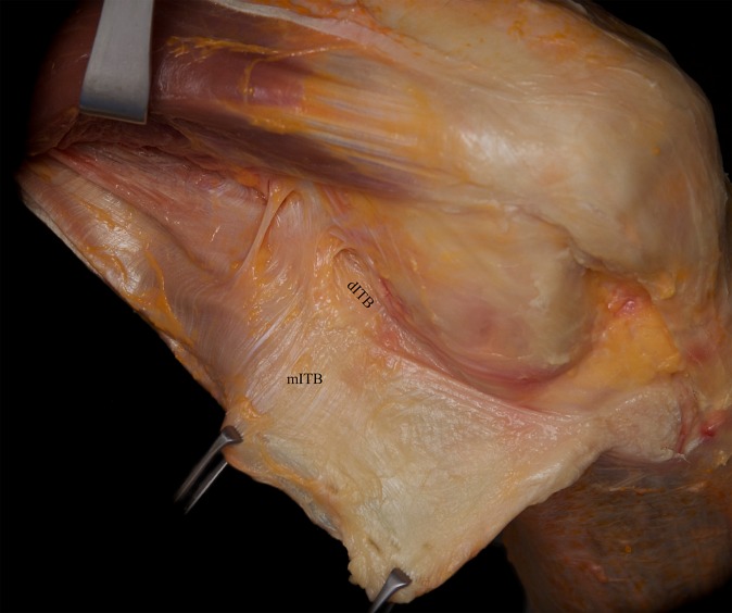
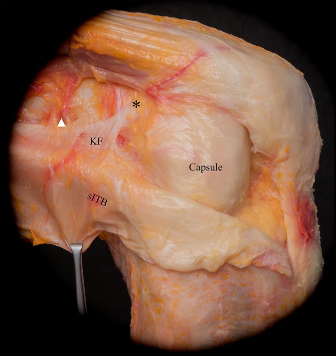
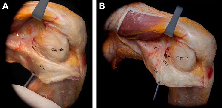
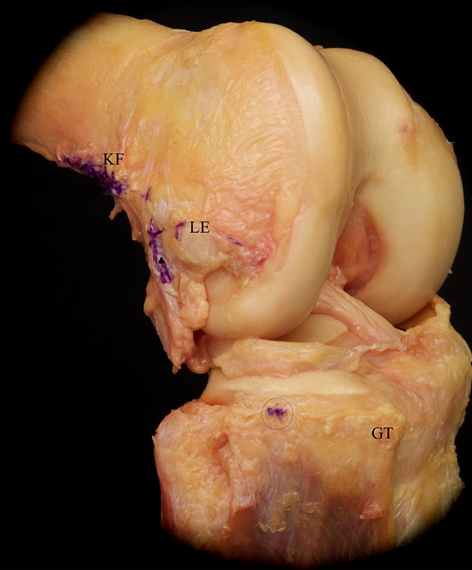
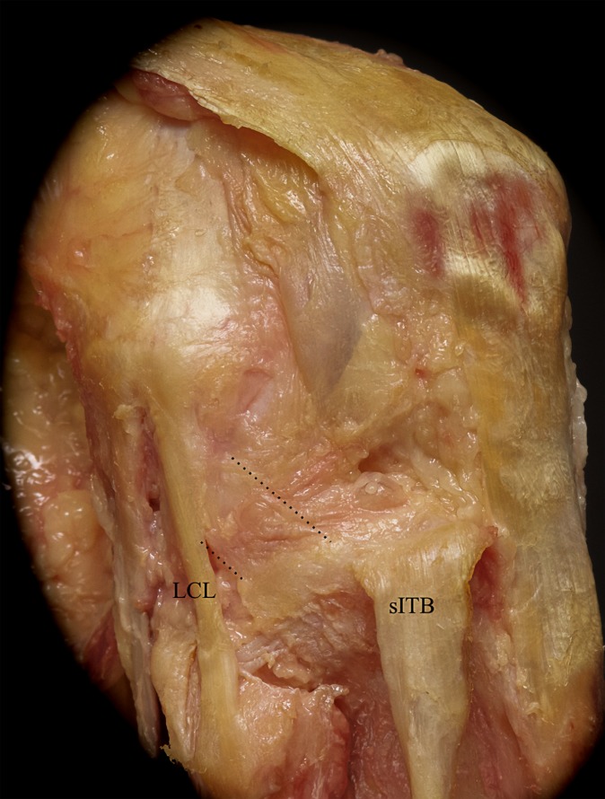
The anterolateral complex of the knee consisted of different distinct layers, with the superficial and deep iliotibial band (ITB) representing layer 1. The superficial ITB had a distinct connection to the distal femoral metaphysis and femoral condyle (Kaplan fibers), and the deep layers of the ITB were identified originating at the level of the Kaplan fibers proximally. This functional unit, consisting of the superficial and deep ITB, was reinforced by the capsulo-osseous layer of the ITB, which was continuous with the fascia of the lateral gastrocnemius and biceps femoris muscles. These 3 components of the ITB became confluent distally, and the insertion spanned from the Gerdy tubercle anteriorly to the lateral tibia posteriorly on a small tubercle (lateral tibial tuberosity). Layer 3 consisted of the anterolateral capsule, in which 35% (7/20) of specimens had a discreet mid-third capsular ligament.
The anterolateral complex consists of the superficial and deep ITB, the capsulo-osseous layer of the ITB, and the anterolateral capsule. The anterolateral complex is defined by the part of the ITB between the Kaplan fibers proximally and its tibial insertion, which forms a functional unit. A discrete anterolateral ligament was not observed; however, the anterolateral ligament described in recent studies likely refers to the capsulo-osseous layer or the mid-third capsular ligament.
The anterolateral knee structures form a complex functional unit. Surgeons should use caution when attempting to restore this intricate structure with extra-articular procedures designed to re-create a single discreet ligament.
关于膝关节前外侧结构存在重大争议。
确定膝关节前外侧复合体的逐层解剖结构。
描述性实验室研究。
对20个新鲜冷冻的尸体膝关节(年龄范围38 - 56岁)进行逐层解剖,以系统地暴露和识别前外侧复合体的各种结构。进行了定量测量,并用高分辨率数字成像记录了每一层。
膝关节前外侧复合体由不同的明显层次组成,浅、深髂胫束(ITB)代表第1层。浅髂胫束与股骨远端干骺端和股骨髁有明显连接(卡普兰纤维),髂胫束深层在近端起源于卡普兰纤维水平。这个由浅、深髂胫束组成的功能单元由髂胫束的关节囊 - 骨膜层加强,该层与腓肠肌外侧头和股二头肌的筋膜连续。髂胫束的这3个组成部分在远端汇合,其止点从前侧的Gerdy结节延伸至后侧胫骨外侧的一个小结节(胫骨外侧结节)。第3层由前外侧关节囊组成,其中35%(7/20)的标本有一条明显的中间三分之一关节囊韧带。
前外侧复合体由浅、深髂胫束、髂胫束的关节囊 - 骨膜层和前外侧关节囊组成。前外侧复合体由近端卡普兰纤维与胫骨止点之间的髂胫束部分所界定,这部分形成一个功能单元。未观察到离散的前外侧韧带;然而,近期研究中描述的前外侧韧带可能指的是关节囊 - 骨膜层或中间三分之一关节囊韧带。
膝关节前外侧结构形成一个复杂的功能单元。外科医生在试图通过旨在重建单一离散韧带的关节外手术来恢复这个复杂结构时应谨慎。