• 文献检索
  • 文档翻译
  • 深度研究
  • 学术资讯
  • Suppr Zotero 插件Zotero 插件
  • 邀请有礼
  • 套餐&价格
  • 历史记录
应用&插件
Suppr Zotero 插件Zotero 插件浏览器插件Mac 客户端Windows 客户端微信小程序
定价
高级版会员购买积分包购买API积分包
服务
文献检索文档翻译深度研究API 文档MCP 服务
关于我们
关于 Suppr公司介绍联系我们用户协议隐私条款
关注我们

Suppr 超能文献

核心技术专利:CN118964589B侵权必究
粤ICP备2023148730 号-1Suppr @ 2026

文献检索

告别复杂PubMed语法,用中文像聊天一样搜索,搜遍4000万医学文献。AI智能推荐,让科研检索更轻松。

立即免费搜索

文件翻译

保留排版,准确专业,支持PDF/Word/PPT等文件格式,支持 12+语言互译。

免费翻译文档

深度研究

AI帮你快速写综述,25分钟生成高质量综述,智能提取关键信息,辅助科研写作。

立即免费体验

小鼠脑血管系统的三维形态学分析:体内和体外方法的比较。

3D morphological analysis of the mouse cerebral vasculature: Comparison of in vivo and ex vivo methods.

作者信息

Steinman Joe, Koletar Margaret M, Stefanovic Bojana, Sled John G

机构信息

Mouse Imaging Centre, The Hospital for Sick Children, Toronto, Ontario, Canada.

Department of Medical Biophysics, University of Toronto, Toronto, Ontario, Canada.

出版信息

PLoS One. 2017 Oct 20;12(10):e0186676. doi: 10.1371/journal.pone.0186676. eCollection 2017.

DOI:10.1371/journal.pone.0186676
PMID:29053753
原文链接:https://pmc.ncbi.nlm.nih.gov/articles/PMC5650181/
Abstract

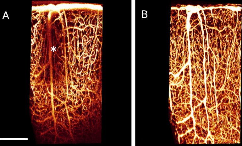
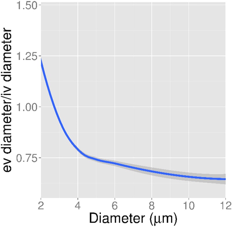
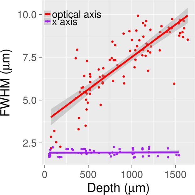
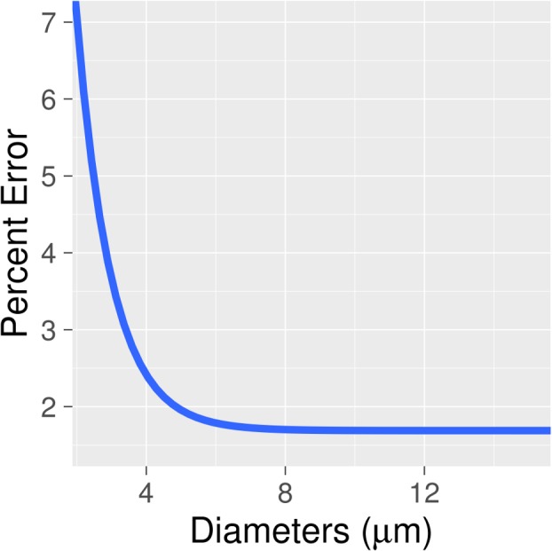
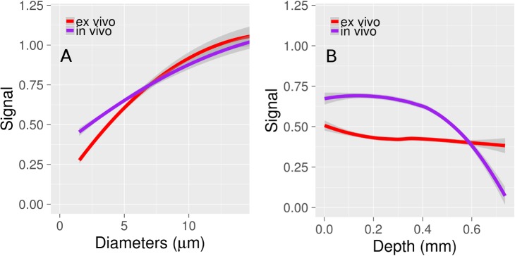
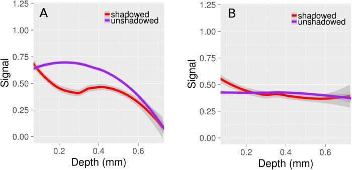
Ex vivo 2-photon fluorescence microscopy (2PFM) with optical clearing enables vascular imaging deep into tissue. However, optical clearing may also produce spherical aberrations if the objective lens is not index-matched to the clearing material, while the perfusion, clearing, and fixation procedure may alter vascular morphology. We compared in vivo and ex vivo 2PFM in mice, focusing on apparent differences in microvascular signal and morphology. Following in vivo imaging, the mice (four total) were perfused with a fluorescent gel and their brains fructose-cleared. The brain regions imaged in vivo were imaged ex vivo. Vessels were segmented in both images using an automated tracing algorithm that accounts for the spatially varying PSF in the ex vivo images. This spatial variance is induced by spherical aberrations caused by imaging fructose-cleared tissue with a water-immersion objective. Alignment of the ex vivo image to the in vivo image through a non-linear warping algorithm enabled comparison of apparent vessel diameter, as well as differences in signal. Shrinkage varied as a function of diameter, with capillaries rendered smaller ex vivo by 13%, while penetrating vessels shrunk by 34%. The pial vasculature attenuated in vivo microvascular signal by 40% 300 μm below the tissue surface, but this effect was absent ex vivo. On the whole, ex vivo imaging was found to be valuable for studying deep cortical vasculature.

摘要

采用光学透明技术的离体双光子荧光显微镜(2PFM)能够对深层组织中的血管进行成像。然而,如果物镜与透明材料的折射率不匹配,光学透明技术也可能产生球差,同时灌注、透明和固定过程可能会改变血管形态。我们比较了小鼠体内和离体2PFM,重点关注微血管信号和形态的明显差异。在体内成像后,对四只小鼠灌注荧光凝胶并对其大脑进行果糖透明处理。对体内成像的脑区进行离体成像。使用自动追踪算法对两张图像中的血管进行分割,该算法考虑了离体图像中空间变化的点扩散函数(PSF)。这种空间变化是由用水浸物镜对果糖透明处理的组织成像所引起的球差导致的。通过非线性扭曲算法将离体图像与体内图像对齐,能够比较表观血管直径以及信号差异。收缩率随直径变化,毛细血管在离体时缩小了13%,而穿通血管缩小了34%。软脑膜血管系统在体内使组织表面以下300μm处的微血管信号衰减了40%,但在离体时这种效应不存在。总体而言,发现离体成像对于研究深层皮质血管系统很有价值。

https://cdn.ncbi.nlm.nih.gov/pmc/blobs/30ea/5650181/b96edcb56d04/pone.0186676.g006.jpg
https://cdn.ncbi.nlm.nih.gov/pmc/blobs/30ea/5650181/7ee90e1fc8bb/pone.0186676.g001.jpg
https://cdn.ncbi.nlm.nih.gov/pmc/blobs/30ea/5650181/5cb767742309/pone.0186676.g002.jpg
https://cdn.ncbi.nlm.nih.gov/pmc/blobs/30ea/5650181/16912cf77a3e/pone.0186676.g003.jpg
https://cdn.ncbi.nlm.nih.gov/pmc/blobs/30ea/5650181/c19502cdc56c/pone.0186676.g004.jpg
https://cdn.ncbi.nlm.nih.gov/pmc/blobs/30ea/5650181/64ee3a52976c/pone.0186676.g005.jpg
https://cdn.ncbi.nlm.nih.gov/pmc/blobs/30ea/5650181/b96edcb56d04/pone.0186676.g006.jpg

相似文献

1
3D morphological analysis of the mouse cerebral vasculature: Comparison of in vivo and ex vivo methods.小鼠脑血管系统的三维形态学分析:体内和体外方法的比较。
PLoS One. 2017 Oct 20;12(10):e0186676. doi: 10.1371/journal.pone.0186676. eCollection 2017.
2
Optical clearing and fluorescence deep-tissue imaging for 3D quantitative analysis of the brain tumor microenvironment.用于脑肿瘤微环境 3D 定量分析的光学透明和荧光深层组织成像。
Angiogenesis. 2017 Nov;20(4):533-546. doi: 10.1007/s10456-017-9565-6. Epub 2017 Jul 11.
3
3D visualization and quantification of microvessels in the whole ischemic mouse brain using solvent-based clearing and light sheet microscopy.使用基于溶剂的透明化方法和光片显微镜对整个缺血小鼠脑内微血管进行三维可视化和定量分析。
J Cereb Blood Flow Metab. 2017 Oct;37(10):3355-3367. doi: 10.1177/0271678X17698970. Epub 2017 Mar 28.
4
Three-dimensional imaging of xenograft tumors using optical computed and emission tomography.利用光学计算机断层扫描和发射断层扫描对异种移植肿瘤进行三维成像。
Med Phys. 2006 Sep;33(9):3193-202. doi: 10.1118/1.2217109.
5
Small molecule fluorophore and copolymer RGD peptide conjugates for ex vivo two-photon fluorescence tumor vasculature imaging.小分子荧光团和共聚物 RGD 肽缀合物用于离体双光子荧光肿瘤血管成像。
Biomaterials. 2012 Nov;33(33):8477-85. doi: 10.1016/j.biomaterials.2012.06.082. Epub 2012 Aug 30.
6
A Simple Technique for Three-Dimensional Imaging and Segmentation of Brain Vasculature U sing Fast Free-of-Acrylamide Clearing Tissue in Murine.一种利用快速无丙烯酰胺清除组织对小鼠脑血管进行三维成像和分割的简单技术。
Cell J. 2019 Apr;21(1):49-56. doi: 10.22074/cellj.2019.5684. Epub 2018 Nov 18.
7
Functional imaging in bulk tissue specimens using optical emission tomography: fluorescence preservation during optical clearing.使用光学发射断层扫描对大块组织标本进行功能成像:光学透明过程中的荧光保存。
Phys Med Biol. 2007 Apr 21;52(8):2035-54. doi: 10.1088/0031-9155/52/8/001. Epub 2007 Mar 20.
8
Optical histology: a method to visualize microvasculature in thick tissue sections of mouse brain.光学组织学:一种可视化小鼠脑厚组织切片中微血管的方法。
PLoS One. 2013;8(1):e53753. doi: 10.1371/journal.pone.0053753. Epub 2013 Jan 23.
9
Automatic anatomical labeling of the complete cerebral vasculature in mouse models.小鼠模型中完整脑血管系统的自动解剖标记
Neuroimage. 2014 Jul 15;95:117-28. doi: 10.1016/j.neuroimage.2014.03.044. Epub 2014 Mar 28.
10
Dynamic contrast-enhanced micro-computed tomography correlates with 3-dimensional fluorescence ultramicroscopy in antiangiogenic therapy of breast cancer xenografts.动态对比增强微计算机断层扫描与三维荧光超显微镜在乳腺癌异种移植抗血管生成治疗中的相关性。
Invest Radiol. 2014 Jul;49(7):445-56. doi: 10.1097/RLI.0000000000000038.

引用本文的文献

1
Deficiency in N-cadherin-Akt3 signaling impairs the blood-brain barrier.N-钙黏蛋白-Akt3信号通路的缺陷会损害血脑屏障。
Cell Rep. 2025 Jun 24;44(6):115831. doi: 10.1016/j.celrep.2025.115831. Epub 2025 Jun 10.
2
Depth-dependent contributions of various vascular zones to cerebral autoregulation and functional hyperemia: An in-silico analysis.不同血管区域对脑自动调节和功能性充血的深度依赖性贡献:一项计算机模拟分析。
PLoS One. 2025 May 19;20(5):e0321053. doi: 10.1371/journal.pone.0321053. eCollection 2025.
3
Deletion of microsomal epoxide hydrolase gene leads to increased density in cerebral vasculature and enhances cerebral blood flow in mice.

本文引用的文献

1
Light-sheet microscopy imaging of a whole cleared rat brain with Thy1-GFP transgene.利用 Thy1-GFP 转基因对整个清除的大鼠脑进行光片显微镜成像。
Sci Rep. 2016 Jun 17;6:28209. doi: 10.1038/srep28209.
2
Mechanical restriction of intracortical vessel dilation by brain tissue sculpts the hemodynamic response.脑组织对皮质内血管扩张的机械性限制塑造了血流动力学反应。
Neuroimage. 2015 Jul 15;115:162-76. doi: 10.1016/j.neuroimage.2015.04.054. Epub 2015 May 5.
3
Pial arteries respond earlier than penetrating arterioles to neural activation in the somatosensory cortex in awake mice exposed to chronic hypoxia: an additional mechanism to proximal integration signaling?
微粒体环氧化物水解酶基因的缺失导致小鼠脑血管密度增加并增强脑血流量。
J Cereb Blood Flow Metab. 2025 Apr 12:271678X251333234. doi: 10.1177/0271678X251333234.
4
Early Activation of Lung CD8 T Cells After Immunization with Live Malaria Sporozoites.用活疟原虫免疫后肺CD8 T细胞的早期激活
Pathog Immun. 2025 Mar 4;10(2):46-68. doi: 10.20411/pai.v10i2.794. eCollection 2025.
5
Non-invasive volumetric ultrasound localization microscopy detects vascular changes in mice with Alzheimer's disease.无创容积超声定位显微镜检测阿尔茨海默病小鼠的血管变化。
Theranostics. 2025 Jan 1;15(3):1110-1121. doi: 10.7150/thno.99097. eCollection 2025.
6
Deep Mouse Brain Two-Photon Near-Infrared Fluorescence Imaging Using a Superconducting Nanowire Single-Photon Detector Array.使用超导纳米线单光子探测器阵列的深度小鼠脑双光子近红外荧光成像
ACS Photonics. 2024 Sep 11;11(10):3960-3971. doi: 10.1021/acsphotonics.4c00111. eCollection 2024 Oct 16.
7
Depth-Dependent Contributions of Various Vascular Zones to Cerebral Autoregulation and Functional Hyperemia: An In-Silico Analysis.不同血管区域对脑自动调节和功能性充血的深度依赖性贡献:一项计算机模拟分析
bioRxiv. 2024 Oct 11:2024.10.07.616950. doi: 10.1101/2024.10.07.616950.
8
Direct association with the vascular basement membrane is a frequent feature of myelinating oligodendrocytes in the neocortex.与血管基底膜的直接关联是新皮层髓鞘少突胶质细胞的一个常见特征。
Fluids Barriers CNS. 2023 Apr 3;20(1):24. doi: 10.1186/s12987-023-00425-4.
9
The post-arteriole transitional zone: a specialized capillary region that regulates blood flow within the CNS microvasculature.后微动脉过渡区:调节中枢神经系统微血管内血流的特殊毛细血管区域。
J Physiol. 2023 Mar;601(5):889-901. doi: 10.1113/JP282246. Epub 2023 Feb 21.
10
Not All Lectins Are Equally Suitable for Labeling Rodent Vasculature.并非所有凝集素都同样适合标记啮齿动物脉管系统。
Int J Mol Sci. 2021 Oct 26;22(21):11554. doi: 10.3390/ijms222111554.
在暴露于慢性缺氧的清醒小鼠中,软脑膜动脉比穿通小动脉对体感皮层的神经激活反应更早:这是近端整合信号的另一种机制吗?
J Cereb Blood Flow Metab. 2014 Nov;34(11):1761-70. doi: 10.1038/jcbfm.2014.140. Epub 2014 Jul 30.
4
Microvascular sprouting, extension, and creation of new capillary connections with adaptation of the neighboring astrocytes in adult mouse cortex under chronic hypoxia.慢性缺氧条件下成年小鼠皮层中小血管发芽、延伸以及新毛细血管连接的形成与邻近星形胶质细胞的适应性变化。
J Cereb Blood Flow Metab. 2014 Feb;34(2):325-31. doi: 10.1038/jcbfm.2013.201. Epub 2013 Nov 20.
5
SeeDB: a simple and morphology-preserving optical clearing agent for neuronal circuit reconstruction.SeeDB:一种简单且保持形态的光透明剂,用于神经元回路重建。
Nat Neurosci. 2013 Aug;16(8):1154-61. doi: 10.1038/nn.3447. Epub 2013 Jun 23.
6
Optical histology: a method to visualize microvasculature in thick tissue sections of mouse brain.光学组织学:一种可视化小鼠脑厚组织切片中微血管的方法。
PLoS One. 2013;8(1):e53753. doi: 10.1371/journal.pone.0053753. Epub 2013 Jan 23.
7
Early-stage microvascular alterations of a new model of controlled cortical traumatic brain injury: 3D morphological analysis using scanning electron microscopy and corrosion casting.采用扫描电子显微镜和腐蚀铸造技术对新型控制性皮质创伤性脑损伤模型的早期微血管改变进行三维形态学分析。
J Neurosurg. 2013 Apr;118(4):763-74. doi: 10.3171/2012.11.JNS12627. Epub 2013 Jan 25.
8
The smallest stroke: occlusion of one penetrating vessel leads to infarction and a cognitive deficit.最小的中风:一支穿支血管闭塞导致梗死和认知障碍。
Nat Neurosci. 2013 Jan;16(1):55-63. doi: 10.1038/nn.3278. Epub 2012 Dec 16.
9
Amyloid-β-dependent compromise of microvascular structure and function in a model of Alzheimer's disease.阿尔茨海默病模型中淀粉样β依赖性的微血管结构和功能损伤。
Brain. 2012 Oct;135(Pt 10):3039-50. doi: 10.1093/brain/aws243.
10
Confocal light sheet microscopy: micron-scale neuroanatomy of the entire mouse brain.共聚焦光片显微镜术:完整小鼠脑的微米级神经解剖结构
Opt Express. 2012 Aug 27;20(18):20582-98. doi: 10.1364/OE.20.020582.