• 文献检索
  • 文档翻译
  • 深度研究
  • 学术资讯
  • Suppr Zotero 插件Zotero 插件
  • 邀请有礼
  • 套餐&价格
  • 历史记录
应用&插件
Suppr Zotero 插件Zotero 插件浏览器插件Mac 客户端Windows 客户端微信小程序
定价
高级版会员购买积分包购买API积分包
服务
文献检索文档翻译深度研究API 文档MCP 服务
关于我们
关于 Suppr公司介绍联系我们用户协议隐私条款
关注我们

Suppr 超能文献

核心技术专利:CN118964589B侵权必究
粤ICP备2023148730 号-1Suppr @ 2026

文献检索

告别复杂PubMed语法,用中文像聊天一样搜索,搜遍4000万医学文献。AI智能推荐,让科研检索更轻松。

立即免费搜索

文件翻译

保留排版,准确专业,支持PDF/Word/PPT等文件格式,支持 12+语言互译。

免费翻译文档

深度研究

AI帮你快速写综述,25分钟生成高质量综述,智能提取关键信息,辅助科研写作。

立即免费体验

替莫唑胺和贝伐单抗治疗后胶质母细胞瘤新生血管模式的改变及其与 DCE-MRI 衍生参数的相关性。

Aberrant glioblastoma neovascularization patterns and their correlation with DCE-MRI-derived parameters following temozolomide and bevacizumab treatment.

机构信息

Department of Radiology, Institute of Surgery Research, Daping Hospital, Third Military Medical University, Chongqing, 400042, China.

Chongqing Clinical Research Center for Imaging and Nuclear Medicine, Chongqing, 400042, China.

出版信息

Sci Rep. 2017 Oct 24;7(1):13894. doi: 10.1038/s41598-017-14341-9.

DOI:10.1038/s41598-017-14341-9
PMID:29066764
原文链接:https://pmc.ncbi.nlm.nih.gov/articles/PMC5654943/
Abstract

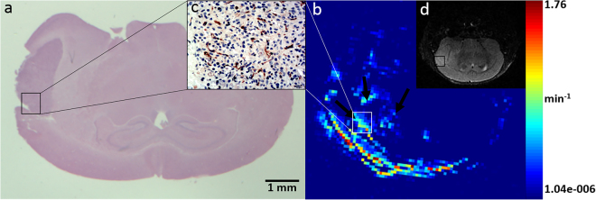
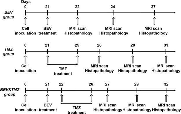
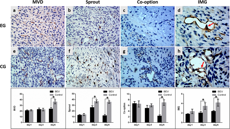
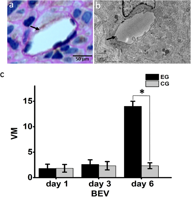
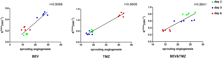
Glioblastoma (GBM) is a highly angiogenic malignancy, and its abundant, aberrant neovascularization is closely related to the proliferation and invasion of tumor cells. However, anti-angiogenesis combined with standard radio-/chemo-therapy produces little improvement in treatment outcomes. Determining the reason for treatment failure is pivotal for GBM treatment. Here, histopathological analysis and dynamic contrast-enhanced MRI (DCE-MRI) were used to explore the effects of temozolomide (TMZ) and bevacizumab (BEV) on GBM neovascularization patterns in an orthotopic U87MG mouse model at 1, 3 and 6 days after treatment. We found that the amount of vascular mimicry (VM) significantly increased 6 days after BEV treatment. TMZ inhibited neovascularization at an early stage, but the microvessel density (MVD) and transfer coefficient (K) derived from DCE-MRI increased 6 days after treatment. TMZ and BEV combination therapy slightly prolonged the inhibitory effect on tumor microvessels. Sprouting angiogenesis was positively correlated with K in all treatment groups. The increase in VM after BEV administration and the increase in MVD and Ktrans after TMZ administration may be responsible for treatment resistance. K holds great potential as an imaging biomarker for indicating the variation in sprouting angiogenesis during drug treatment for GBM.

摘要

胶质母细胞瘤(GBM)是一种高度血管生成的恶性肿瘤,其丰富的、异常的新生血管化与肿瘤细胞的增殖和浸润密切相关。然而,抗血管生成联合标准的放化疗并没有明显改善治疗效果。确定治疗失败的原因对于 GBM 的治疗至关重要。在这里,我们使用组织病理学分析和动态对比增强 MRI(DCE-MRI),在治疗后 1、3 和 6 天,在原位 U87MG 小鼠模型中,探索替莫唑胺(TMZ)和贝伐单抗(BEV)对 GBM 新生血管模式的影响。我们发现,BEV 治疗 6 天后血管模拟(VM)的数量显著增加。TMZ 在早期抑制新生血管形成,但 DCE-MRI 得出的微血管密度(MVD)和转移系数(K)在治疗后 6 天增加。TMZ 和 BEV 联合治疗略微延长了对肿瘤微血管的抑制作用。所有治疗组中,发芽型血管生成与 K 呈正相关。BEV 给药后 VM 的增加以及 TMZ 给药后 MVD 和 Ktrans 的增加,可能是导致治疗耐药的原因。K 作为一种成像生物标志物,具有很大的潜力,可以指示 GBM 药物治疗期间发芽型血管生成的变化。

https://cdn.ncbi.nlm.nih.gov/pmc/blobs/9f55/5654943/18dd5ecbe5b1/41598_2017_14341_Fig8_HTML.jpg
https://cdn.ncbi.nlm.nih.gov/pmc/blobs/9f55/5654943/dc52863e95c4/41598_2017_14341_Fig1_HTML.jpg
https://cdn.ncbi.nlm.nih.gov/pmc/blobs/9f55/5654943/bbe39da22dfc/41598_2017_14341_Fig2_HTML.jpg
https://cdn.ncbi.nlm.nih.gov/pmc/blobs/9f55/5654943/f0658f89495f/41598_2017_14341_Fig3_HTML.jpg
https://cdn.ncbi.nlm.nih.gov/pmc/blobs/9f55/5654943/73cf7acc7e9e/41598_2017_14341_Fig4_HTML.jpg
https://cdn.ncbi.nlm.nih.gov/pmc/blobs/9f55/5654943/48077272b5fd/41598_2017_14341_Fig5_HTML.jpg
https://cdn.ncbi.nlm.nih.gov/pmc/blobs/9f55/5654943/26c5cf628024/41598_2017_14341_Fig6_HTML.jpg
https://cdn.ncbi.nlm.nih.gov/pmc/blobs/9f55/5654943/5bf7d9b5d1e1/41598_2017_14341_Fig7_HTML.jpg
https://cdn.ncbi.nlm.nih.gov/pmc/blobs/9f55/5654943/18dd5ecbe5b1/41598_2017_14341_Fig8_HTML.jpg

相似文献

1
Aberrant glioblastoma neovascularization patterns and their correlation with DCE-MRI-derived parameters following temozolomide and bevacizumab treatment.替莫唑胺和贝伐单抗治疗后胶质母细胞瘤新生血管模式的改变及其与 DCE-MRI 衍生参数的相关性。
Sci Rep. 2017 Oct 24;7(1):13894. doi: 10.1038/s41598-017-14341-9.
2
Dual inhibition of PFKFB3 and VEGF normalizes tumor vasculature, reduces lactate production, and improves chemotherapy in glioblastoma: insights from protein expression profiling and MRI.双重抑制 PFKFB3 和 VEGF 可使肿瘤血管正常化,减少乳酸生成,并改善胶质母细胞瘤的化疗效果:来自蛋白表达谱和 MRI 的见解。
Theranostics. 2020 Jun 5;10(16):7245-7259. doi: 10.7150/thno.44427. eCollection 2020.
3
Amino acid PET tracers are reliable markers of treatment responses to single-agent or combination therapies including temozolomide, interferon-β, and/or bevacizumab for glioblastoma.氨基酸正电子发射断层显像(PET)示踪剂是胶质母细胞瘤对包括替莫唑胺、干扰素-β和/或贝伐单抗在内的单药或联合治疗反应的可靠标志物。
Nucl Med Biol. 2015 Jul;42(7):598-607. doi: 10.1016/j.nucmedbio.2015.01.008. Epub 2015 Jan 31.
4
[Ga]RGD Versus [F]FDG PET Imaging in Monitoring Treatment Response of a Mouse Model of Human Glioblastoma Tumor with Bevacizumab and/or Temozolomide.[Ga]RGD 与 [F]FDG PET 成像在贝伐单抗和/或替莫唑胺治疗人胶质母细胞瘤肿瘤模型中的应用。
Mol Imaging Biol. 2019 Apr;21(2):297-305. doi: 10.1007/s11307-018-1224-9.
5
First-line bevacizumab contributes to survival improvement in glioblastoma patients complementary to temozolomide.贝伐珠单抗联合替莫唑胺一线治疗胶质母细胞瘤可改善患者生存。
J Neurooncol. 2020 Feb;146(3):451-458. doi: 10.1007/s11060-019-03339-0. Epub 2020 Feb 4.
6
Noninvasive Characterization of Tumor Angiogenesis and Oxygenation in Bevacizumab-treated Recurrent Glioblastoma by Using Dynamic Susceptibility MRI: Secondary Analysis of the European Organization for Research and Treatment of Cancer 26101 Trial.使用动态磁敏感对比 MRI 对贝伐珠单抗治疗后复发性脑胶质瘤的肿瘤血管生成和氧合状态进行无创性特征描述:欧洲癌症研究与治疗组织 26101 试验的二次分析。
Radiology. 2020 Oct;297(1):164-175. doi: 10.1148/radiol.2020200978. Epub 2020 Jul 28.
7
Mechanistic interrogation of combination bevacizumab/dual PI3K/mTOR inhibitor response in glioblastoma implementing novel MR and PET imaging biomarkers.使用新型磁共振成像(MR)和正电子发射断层显像(PET)生物标志物对胶质母细胞瘤中贝伐单抗/双磷脂酰肌醇-3激酶(PI3K)/雷帕霉素靶蛋白(mTOR)抑制剂联合治疗反应进行机制研究。
Eur J Nucl Med Mol Imaging. 2016 Aug;43(9):1673-83. doi: 10.1007/s00259-016-3343-3. Epub 2016 Mar 15.
8
Decreased APE-1 by Nitroxoline Enhances Therapeutic Effect in a Temozolomide-resistant Glioblastoma: Correlation with Diffusion Weighted Imaging.硝呋莫司降低 APE-1 表达增强替莫唑胺耐药脑胶质瘤治疗效果:与弥散加权成像的相关性。
Sci Rep. 2019 Nov 12;9(1):16613. doi: 10.1038/s41598-019-53147-9.
9
Proteasome inhibition with bortezomib induces cell death in GBM stem-like cells and temozolomide-resistant glioma cell lines, but stimulates GBM stem-like cells' VEGF production and angiogenesis.硼替佐米抑制蛋白酶体诱导 GBM 干细胞样细胞和替莫唑胺耐药神经胶质瘤细胞系的细胞死亡,但刺激 GBM 干细胞样细胞的 VEGF 产生和血管生成。
J Neurosurg. 2013 Dec;119(6):1415-23. doi: 10.3171/2013.7.JNS1323. Epub 2013 Oct 4.
10
Survival benefit of glioblastoma patients after FDA approval of temozolomide concomitant with radiation and bevacizumab: A population-based study.美国食品药品监督管理局批准替莫唑胺联合放疗和贝伐单抗后胶质母细胞瘤患者的生存获益:一项基于人群的研究。
Oncotarget. 2017 Jul 4;8(27):44015-44031. doi: 10.18632/oncotarget.17054.

引用本文的文献

1
The Roles of RNA-Binding Proteins in Vasculogenic Mimicry Regulation in Glioblastoma.RNA结合蛋白在胶质母细胞瘤血管生成拟态调节中的作用
Int J Mol Sci. 2025 Aug 18;26(16):7976. doi: 10.3390/ijms26167976.
2
Role of dynamic contrast-enhanced and dynamic susceptibility contrast imaging in evaluating the biological features of glioma.动态对比增强成像和动态磁敏感对比成像在评估胶质瘤生物学特征中的作用。
Quant Imaging Med Surg. 2025 Aug 1;15(8):7030-7045. doi: 10.21037/qims-2024-2794. Epub 2025 Jul 28.
3
Dynamic Contrast-enhanced MRI Processing Comparison for Distinguishing True Progression From Pseudoprogression in High-grade Glioma.

本文引用的文献

1
Evaluation of neovascularization patterns in an orthotopic rat glioma model with dynamic contrast-enhanced MRI.利用动态对比增强磁共振成像评估原位大鼠胶质瘤模型中的新生血管形成模式
Acta Radiol. 2017 Sep;58(9):1138-1146. doi: 10.1177/0284185116681038. Epub 2016 Dec 12.
2
MR Imaging Biomarkers to Monitor Early Response to Hypoxia-Activated Prodrug TH-302 in Pancreatic Cancer Xenografts.用于监测胰腺癌异种移植模型中对缺氧激活前药TH-302早期反应的磁共振成像生物标志物
PLoS One. 2016 May 26;11(5):e0155289. doi: 10.1371/journal.pone.0155289. eCollection 2016.
3
Galunisertib inhibits glioma vasculogenic mimicry formation induced by astrocytes.
动态对比增强磁共振成像处理在高级别胶质瘤中区分真性进展与假性进展的比较
J Comput Assist Tomogr. 2025;49(4):656-661. doi: 10.1097/RCT.0000000000001716. Epub 2025 Jan 27.
4
Galunisertib promotes bevacizumab-induced vascular normalization in nasopharyngeal carcinoma: Multi-parameter MRI evaluation.加芦米司特促进贝伐单抗诱导的鼻咽癌血管正常化:多参数MRI评估
Mol Ther Oncol. 2024 Aug 8;32(3):200858. doi: 10.1016/j.omton.2024.200858. eCollection 2024 Sep 19.
5
The in vitro dynamics of pseudo-vascular network formation.体外伪血管网络形成的动力学。
Br J Cancer. 2024 Aug;131(3):457-467. doi: 10.1038/s41416-024-02722-7. Epub 2024 Jun 20.
6
HNRNPA2B1 stabilizes NFATC3 levels to potentiate its combined actions with FOSL1 to mediate vasculogenic mimicry in GBM cells.HNRNPA2B1 稳定 NFATC3 水平,增强其与 FOSL1 的协同作用,从而介导 GBM 细胞中的血管生成拟态。
Cell Biol Toxicol. 2024 Jun 11;40(1):44. doi: 10.1007/s10565-024-09890-5.
7
Flexible Toolbox of High-Precision Microfluidic Modules for Versatile Droplet-Based Applications.用于基于微滴的多功能应用的高精度微流控模块灵活工具箱
Micromachines (Basel). 2024 Feb 7;15(2):250. doi: 10.3390/mi15020250.
8
Predictive value of MRI features on glioblastoma.胶质母细胞瘤MRI特征的预测价值
Eur Radiol. 2023 Jun;33(6):4472-4474. doi: 10.1007/s00330-023-09535-x. Epub 2023 Apr 5.
9
Differential P-Glycoprotein/CD31 Expression as Markers of Vascular Co-Option in Primary Central Nervous System Tumors.差异表达的P-糖蛋白/CD31作为原发性中枢神经系统肿瘤血管共选的标志物
Diagnostics (Basel). 2022 Dec 10;12(12):3120. doi: 10.3390/diagnostics12123120.
10
Glioblastoma-Specific Strategies of Vascularization: Implications in Anti-Angiogenic Therapy Resistance.胶质母细胞瘤特异性血管生成策略:对抗血管生成治疗耐药性的影响
J Pers Med. 2022 Oct 1;12(10):1625. doi: 10.3390/jpm12101625.
加鲁尼西替布可抑制星形胶质细胞诱导的胶质瘤血管生成拟态形成。
Sci Rep. 2016 Mar 15;6:23056. doi: 10.1038/srep23056.
4
Angiogenesis Revisited: An Overlooked Role of Endothelial Cell Metabolism in Vessel Sprouting.重新审视血管生成:内皮细胞代谢在血管萌芽中被忽视的作用
Microcirculation. 2015 Oct;22(7):509-17. doi: 10.1111/micc.12229.
5
Anti-angiogenic Effects of Bumetanide Revealed by DCE-MRI with a Biodegradable Macromolecular Contrast Agent in a Colon Cancer Model.在结肠癌模型中,使用可生物降解的大分子造影剂通过动态对比增强磁共振成像揭示布美他尼的抗血管生成作用。
Pharm Res. 2015 Sep;32(9):3029-43. doi: 10.1007/s11095-015-1684-4. Epub 2015 Apr 4.
6
Cluster analysis of quantitative parametric maps from DCE-MRI: application in evaluating heterogeneity of tumor response to antiangiogenic treatment.基于动态对比增强磁共振成像(DCE-MRI)定量参数图的聚类分析:在评估肿瘤抗血管生成治疗反应异质性中的应用
Magn Reson Imaging. 2015 Jul;33(6):725-36. doi: 10.1016/j.mri.2015.03.005. Epub 2015 Mar 31.
7
Glipizide suppresses embryonic vasculogenesis and angiogenesis through targeting natriuretic peptide receptor A.格列吡嗪通过靶向利钠肽受体A抑制胚胎血管生成和血管新生。
Exp Cell Res. 2015 May 1;333(2):261-272. doi: 10.1016/j.yexcr.2015.03.012. Epub 2015 Mar 28.
8
MicroRNA-184 modulates canonical Wnt signaling through the regulation of frizzled-7 expression in the retina with ischemia-induced neovascularization.微小RNA-184通过调控视网膜中卷曲蛋白-7的表达来调节经典Wnt信号通路,该视网膜存在缺血诱导的新生血管形成。
FEBS Lett. 2015 Apr 28;589(10):1143-1149. doi: 10.1016/j.febslet.2015.03.010. Epub 2015 Mar 18.
9
Diagnosis of brain tumors using dynamic contrast-enhanced perfusion imaging with a short acquisition time.使用短采集时间的动态对比增强灌注成像诊断脑肿瘤。
Springerplus. 2015 Feb 24;4:88. doi: 10.1186/s40064-015-0861-6. eCollection 2015.
10
Successful combination of sunitinib and girentuximab in two renal cell carcinoma animal models: a rationale for combination treatment of patients with advanced RCC.舒尼替尼与吉伦特单抗在两种肾细胞癌动物模型中的成功联合:晚期肾细胞癌患者联合治疗的理论依据
Neoplasia. 2015 Feb;17(2):215-24. doi: 10.1016/j.neo.2014.12.011.