Tiede-Lewis LeAnn M, Xie Yixia, Hulbert Molly A, Campos Richard, Dallas Mark R, Dusevich Vladimir, Bonewald Lynda F, Dallas Sarah L
Department of Oral and Craniofacial Sciences, School of Dentistry, University of Missouri Kansas City, Kansas City, MO 64108, USA.
Departments of Anatomy and Cell Biology and Orthopaedic Surgery, School of Medicine, Indiana University, Indianapolis, IN 46202, USA.
Aging (Albany NY). 2017 Oct 26;9(10):2190-2208. doi: 10.18632/aging.101308.
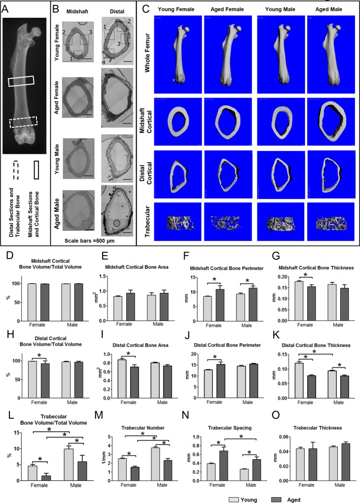
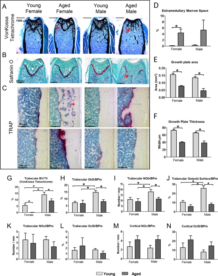
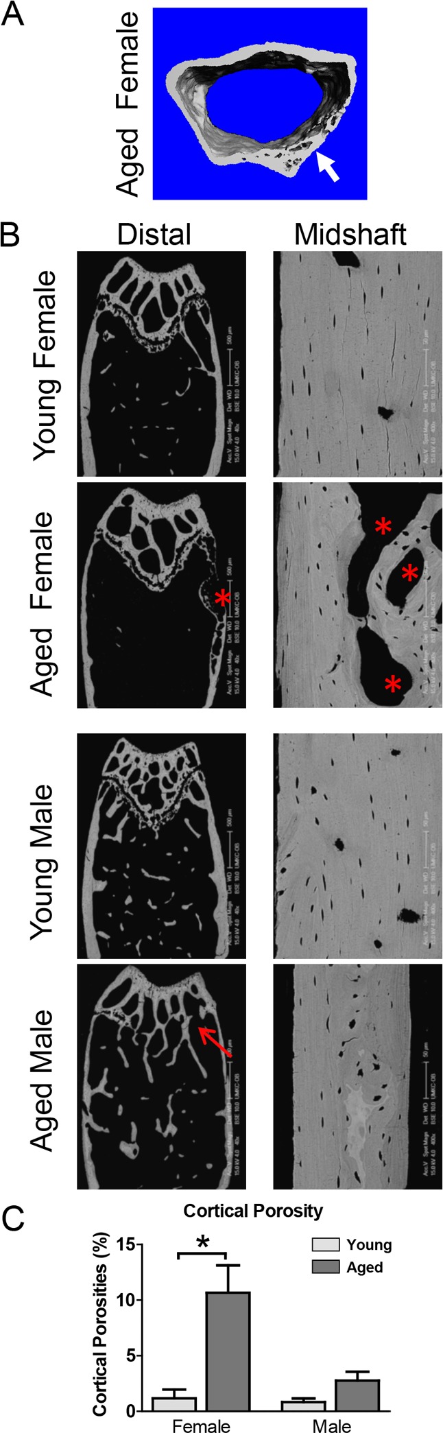
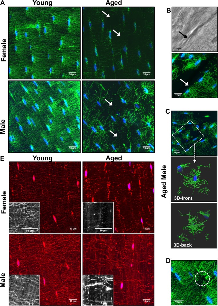
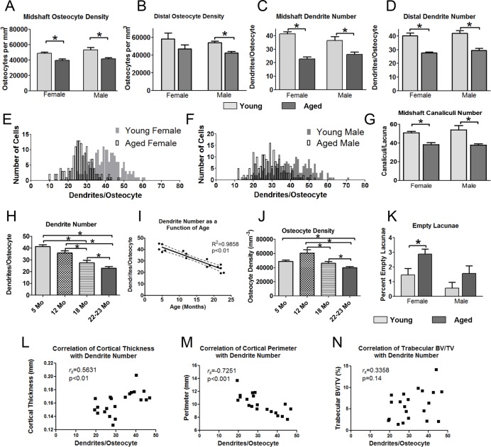
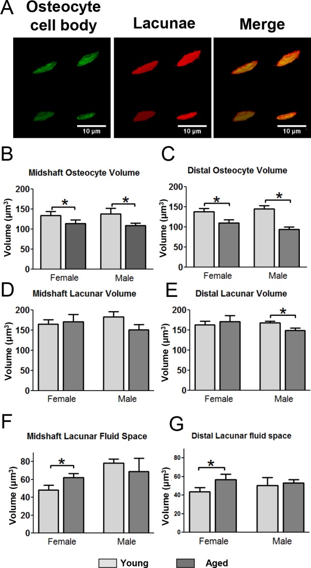
Age-related bone loss and associated fracture risk are major problems in musculoskeletal health. Osteocytes have emerged as key regulators of bone mass and as a therapeutic target for preventing bone loss. As aging is associated with changes in the osteocyte lacunocanalicular system, we focused on the responsible cellular mechanisms in osteocytes. Bone phenotypic analysis was performed in young-(5mo) and aged-(22mo) C57BL/6 mice and changes in bone structure/geometry correlated with alterations in osteocyte parameters determined using novel multiplexed-3D-confocal imaging techniques. Age-related bone changes analogous to those in humans were observed, including increased cortical diameter, decreased cortical thickness, reduced trabecular BV/TV and cortical porosities. This was associated with a dramatic reduction in osteocyte dendrite number and cell density, particularly in females, where osteocyte dendricity decreased linearly from 5, 12, 18 to 22mo and correlated significantly with cortical bone parameters. Reduced dendricity preceded decreased osteocyte number, suggesting dendrite loss may trigger loss of viability. Age-related degeneration of osteocyte networks may impair bone anabolic responses to loading and gender differences in osteocyte cell body and lacunar fluid volumes we observed in aged mice may lead to gender-related differences in mechanosensitivity. Therapies to preserve osteocyte dendricity and viability may be beneficial for bone health in aging.
与年龄相关的骨质流失及相关骨折风险是肌肉骨骼健康领域的主要问题。骨细胞已成为骨量的关键调节因子以及预防骨质流失的治疗靶点。由于衰老与骨细胞腔隙小管系统的变化有关,我们聚焦于骨细胞中相关的细胞机制。对年轻(5月龄)和老龄(22月龄)的C57BL/6小鼠进行了骨表型分析,并使用新型多重三维共聚焦成像技术确定骨细胞参数的变化与骨结构/几何形态的改变相关。观察到了与人类相似的与年龄相关的骨骼变化,包括皮质直径增加、皮质厚度减小、小梁骨体积分数降低以及皮质孔隙率增加。这与骨细胞树突数量和细胞密度的显著降低有关,尤其是在雌性小鼠中,骨细胞树突从5、12、18月龄到22月龄呈线性下降,并与皮质骨参数显著相关。树突减少先于骨细胞数量减少,提示树突丧失可能引发活力丧失。骨细胞网络的与年龄相关的退化可能会损害骨骼对负荷的合成代谢反应,并且我们在老龄小鼠中观察到的骨细胞胞体和腔隙液体积的性别差异可能导致机械敏感性的性别差异。保留骨细胞树突和活力的疗法可能有益于衰老过程中的骨骼健康。