Medical Clinic I, University Hospital Carl Gustav Carus Dresden, Heidelberg, Germany.
Medical Clinic III, University Hospital Carl Gustav Carus Dresden, Heidelberg, Germany.
Haematologica. 2018 Jan;103(1):61-68. doi: 10.3324/haematol.2017.172726. Epub 2017 Oct 27.
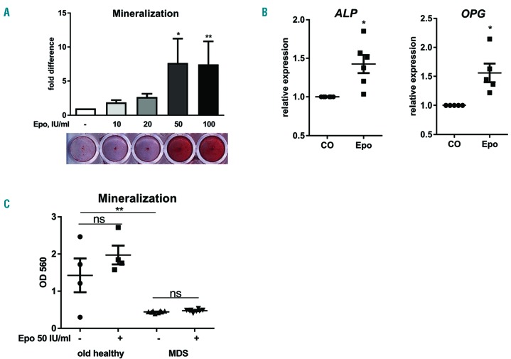
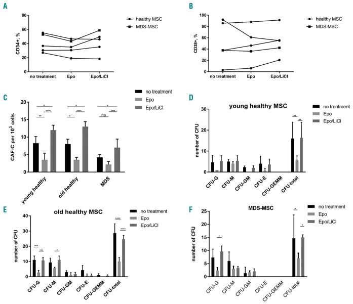
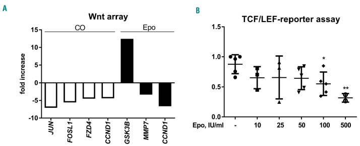
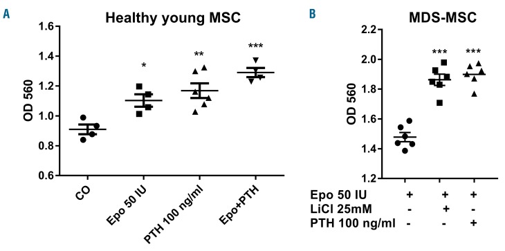
The effects of erythropoietin on osteoblasts and bone formation are controversial. Since patients with myelodysplastic syndromes often display excessively high erythropoietin levels, we aimed to analyze the effect of erythropoietin on osteoblast function in myelodysplastic syndromes and define the role of Wnt signaling in this process. Expression of osteoblast-specific genes and subsequent osteoblast mineralization was increased in mesenchymal stromal cells from healthy young donors by erythropoietin treatment. However, erythropoietin failed to increase osteoblast mineralization in old healthy donors and in patients with myelodysplasia, whereas the basal differentiation potential of the latter was already significantly reduced compared to that of age-matched controls (<0.01). This was accompanied by a significantly reduced expression of genes of the canonical Wnt pathway. Treatment of these cells with erythropoietin further inhibited the canonical Wnt pathway. Exposure of murine cells (C2C12) to erythropoietin also produced a dose-dependent inhibition of TCF/LEF promoter activity (maximum at 500 IU/mL, -2.8-fold; <0.01). The decreased differentiation capacity of erythropoietin-pretreated mesenchymal stromal cells from patients with myelodysplasia could be restored by activating the Wnt pathway using lithium chloride or parathyroid hormone. Its hematopoiesis-supporting capacity was reduced, while reactivation of the canonical Wnt pathway in mesenchymal stromal cells could reverse this effect. Thus, these data demonstrate that erythropoietin modulates components of the osteo-hematopoietic niche in a context-dependent manner being anabolic in young, but catabolic in mature bone cells. Targeting the Wnt pathway in patients with myelodysplastic syndromes may be an appealing strategy to promote the functional capacity of the osteo-hematopoietic niche.
促红细胞生成素对成骨细胞和骨形成的影响存在争议。由于骨髓增生异常综合征患者通常表现出过高的促红细胞生成素水平,我们旨在分析促红细胞生成素对骨髓增生异常综合征中成骨细胞功能的影响,并确定 Wnt 信号通路在这一过程中的作用。促红细胞生成素处理可增加健康年轻供体间充质基质细胞中成骨细胞特异性基因的表达,并随后增加成骨细胞矿化。然而,促红细胞生成素未能增加老年健康供体和成骨髓增生异常患者的成骨细胞矿化,而后者的基础分化潜力已明显低于年龄匹配的对照组(<0.01)。这伴随着经典 Wnt 通路基因表达的显著减少。用促红细胞生成素处理这些细胞进一步抑制了经典 Wnt 通路。促红细胞生成素暴露于鼠细胞(C2C12)也产生了 TCF/LEF 启动子活性的剂量依赖性抑制(最大为 500 IU/mL,-2.8 倍;<0.01)。骨髓增生异常患者预处理间充质基质细胞分化能力的降低可通过使用氯化锂或甲状旁腺激素激活 Wnt 通路来恢复。其造血支持能力降低,而间充质基质细胞中经典 Wnt 通路的再激活可以逆转这种效应。因此,这些数据表明,促红细胞生成素以依赖于上下文的方式调节造血龛的组成部分,在年轻的成骨细胞中是合成代谢的,但在成熟的成骨细胞中是分解代谢的。针对骨髓增生异常综合征患者的 Wnt 通路可能是一种有吸引力的策略,可促进造血龛的功能能力。