• 文献检索
  • 文档翻译
  • 深度研究
  • 学术资讯
  • Suppr Zotero 插件Zotero 插件
  • 邀请有礼
  • 套餐&价格
  • 历史记录
应用&插件
Suppr Zotero 插件Zotero 插件浏览器插件Mac 客户端Windows 客户端微信小程序
定价
高级版会员购买积分包购买API积分包
服务
文献检索文档翻译深度研究API 文档MCP 服务
关于我们
关于 Suppr公司介绍联系我们用户协议隐私条款
关注我们

Suppr 超能文献

核心技术专利:CN118964589B侵权必究
粤ICP备2023148730 号-1Suppr @ 2026

文献检索

告别复杂PubMed语法,用中文像聊天一样搜索,搜遍4000万医学文献。AI智能推荐,让科研检索更轻松。

立即免费搜索

文件翻译

保留排版,准确专业,支持PDF/Word/PPT等文件格式,支持 12+语言互译。

免费翻译文档

深度研究

AI帮你快速写综述,25分钟生成高质量综述,智能提取关键信息,辅助科研写作。

立即免费体验

数字化口腔内X光片灰度与CT扫描亨氏单位之间关系的体外评估

In vitro Evaluation of the Relationship between Gray Scales in Digital Intraoral Radiographs and Hounsfield Units in CT Scans.

作者信息

Khojastepour L, Mohammadzadeh S, Jazayeri M, Omidi M

机构信息

Professor of Oral and Maxillofacial Radiology (M.Sc.), Department of Radiology, School of Dentistry, Shiraz University of Medical Sciences, Shiraz, Iran.

Specialist in Periodontology (M.Sc.), Department of Periodontics, School of Dentistry, Bushehr University of Medical Sciences, Bushehr, Iran.

出版信息

J Biomed Phys Eng. 2017 Sep 1;7(3):289-298. eCollection 2017 Sep.

PMID:29082220
原文链接:https://pmc.ncbi.nlm.nih.gov/articles/PMC5654135/
Abstract

BACKGROUND

Jaw bone quality plays an essential role in treatment planning and prognosis of dental implants. Regarding several available methods for bone density measurements, they are not routinely used before implant surgery due to hard accessibility.

OBJECTIVE

An in vitro investigation of correlation between average gray scale in direct digital radiographs and Hounsfield units in CT-Scan provides a feasible method for evaluating alveolar bone quality prior to implant surgery.

METHODS

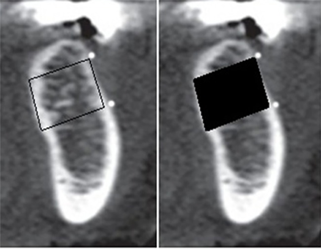
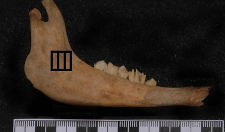
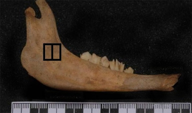
26 sheep's mandibles in which a square shape ROI marked by gutta percha, were prepared. Three direct digital radiographs (CCD sensor) from every specimen were taken using 80, 100 and 200 milli-seconds. Then, the average gray levels for ROIs were calculated using a costume-made software. Next, the specimens were scanned using a 16-slice spiral CT and the Hounsfield Unit of each ROI was calculated. Pearson analysis measured the correlation between Hounsfield units and average gray levels.

RESULTS

There was a positive correlation between Hounsfield unit and average gray level in the radiographs and the correlation was better in higher exposure times.

CONCLUSION

It is possible to estimate Hounsfield unit and bone density in the jaw bones using average gray scale in a digital radiograph. This approach is easy, simple and available and also results in lower patient exposure comparing other bone densitometric analysis methods.

摘要

背景

颌骨质量在牙种植体治疗计划和预后中起着至关重要的作用。关于几种现有的骨密度测量方法,由于难以获取,在种植手术前并未常规使用。

目的

对直接数字化X线片的平均灰度与CT扫描中的亨氏单位之间的相关性进行体外研究,为种植手术前评估牙槽骨质量提供一种可行的方法。

方法

准备26个用牙胶标记出方形感兴趣区(ROI)的绵羊下颌骨。使用80、100和200毫秒对每个标本拍摄三张直接数字化X线片(CCD传感器)。然后,使用定制软件计算ROI的平均灰度。接下来,使用16层螺旋CT对标本进行扫描,并计算每个ROI的亨氏单位。Pearson分析测量亨氏单位与平均灰度之间的相关性。

结果

X线片中亨氏单位与平均灰度之间存在正相关,且在较高曝光时间下相关性更好。

结论

利用数字化X线片中的平均灰度来估计颌骨中的亨氏单位和骨密度是可行的。这种方法简便易行且可获得,与其他骨密度分析方法相比,还能减少患者的辐射暴露。

https://cdn.ncbi.nlm.nih.gov/pmc/blobs/4699/5654135/84ce9d4fef71/JBPE-7-289-g004.jpg
https://cdn.ncbi.nlm.nih.gov/pmc/blobs/4699/5654135/40de1fa64fcc/JBPE-7-289-g001.jpg
https://cdn.ncbi.nlm.nih.gov/pmc/blobs/4699/5654135/5e4cd937fa05/JBPE-7-289-g002.jpg
https://cdn.ncbi.nlm.nih.gov/pmc/blobs/4699/5654135/60c8f76d54c6/JBPE-7-289-g003.jpg
https://cdn.ncbi.nlm.nih.gov/pmc/blobs/4699/5654135/84ce9d4fef71/JBPE-7-289-g004.jpg

相似文献

1
In vitro Evaluation of the Relationship between Gray Scales in Digital Intraoral Radiographs and Hounsfield Units in CT Scans.数字化口腔内X光片灰度与CT扫描亨氏单位之间关系的体外评估
J Biomed Phys Eng. 2017 Sep 1;7(3):289-298. eCollection 2017 Sep.
2
Relationship between Hounsfield Unit in CT Scan and Gray Scale in CBCT.CT扫描中的亨氏单位与CBCT中的灰度之间的关系。
J Dent Res Dent Clin Dent Prospects. 2014 Spring;8(2):107-10. doi: 10.5681/joddd.2014.019. Epub 2014 Jun 11.
3
Use of digital panoramic radiology in presurgical implant treatment planning to accurately assess bone density.
J Prosthet Dent. 2016 Aug;116(2):200-205.e1. doi: 10.1016/j.prosdent.2016.01.017. Epub 2016 Mar 30.
4
Comparison of the Hounsfield unit in CT scan with the gray level in cone-beam CT.CT扫描中的亨氏单位与锥形束CT中的灰度对比。
J Dent Res Dent Clin Dent Prospects. 2019 Summer;13(3):177-182. doi: 10.15171/joddd.2019.028. Epub 2019 Oct 7.
5
Conventional multi-slice computed tomography (CT) and cone-beam CT (CBCT) for computer-assisted implant placement. Part I: relationship of radiographic gray density and implant stability.用于计算机辅助种植体植入的传统多层螺旋计算机断层扫描(CT)和锥束CT(CBCT)。第一部分:影像学灰度密度与种植体稳定性的关系。
Clin Implant Dent Relat Res. 2013 Dec;15(6):893-906. doi: 10.1111/j.1708-8208.2011.00436.x. Epub 2012 Jan 17.
6
Bone quality evaluation at dental implant site using multislice CT, micro-CT, and cone beam CT.使用多层螺旋CT、显微CT和锥形束CT对牙种植部位进行骨质量评估。
Clin Oral Implants Res. 2015;26(1):e1-7. doi: 10.1111/clr.12315. Epub 2013 Dec 11.
7
Evaluation of a Novel Cone Beam Computed Tomography Scanner for Bone Density Examinations in Preoperative 3D Reconstructions and Correlation with Primary Implant Stability.一种新型锥形束计算机断层扫描仪在术前三维重建中进行骨密度检查及与种植体初期稳定性相关性的评估。
Clin Implant Dent Relat Res. 2015 Oct;17(5):844-53. doi: 10.1111/cid.12193. Epub 2013 Dec 27.
8
Implant Stability and Bone Characteristics in Free Fibula Flaps Used for Jaw Reconstruction: A Prospective Cohort Study.
Int J Oral Maxillofac Implants. 2017 September/October;32(5):1145–1152. doi: 10.11607/jomi.5359. Epub 2017 Mar 17.
9
Computer-assisted densitometric image analysis of digital subtraction images: in vivo error of the method and effect of thresholding.数字减影图像的计算机辅助密度测量图像分析:该方法的体内误差及阈值化的影响。
J Periodontol. 1998 Sep;69(9):967-74. doi: 10.1902/jop.1998.69.9.967.
10
Quantitative evaluation of bone density using the Hounsfield index.使用亨氏单位指数对骨密度进行定量评估。
Int J Oral Maxillofac Implants. 2006 Mar-Apr;21(2):290-7.

引用本文的文献

1
A New Method for the Digital Assessment of the Relative Density of Bone Tissue in Dentistry Using the ImageJ Software Package.一种使用ImageJ软件包对牙科中骨组织相对密度进行数字评估的新方法。
Dent J (Basel). 2025 Aug 19;13(8):375. doi: 10.3390/dj13080375.
2
Assessment of CBCT gray value in different regions-of-interest and fields-of-view compared to Hounsfield unit.评估不同感兴趣区和视野下的 CBCT 灰度值与亨氏单位的关系。
Dentomaxillofac Radiol. 2023 Nov;52(8):20230187. doi: 10.1259/dmfr.20230187. Epub 2023 Oct 24.
3
Analysis of Bone Density and Bone Morphometry by Periapical Radiographs in Dental Implant Osseointegration Process.

本文引用的文献

1
Validity of wax and acrylic as soft-tissue simulation materials used in in vitro radiographic studies.蜡和丙烯酸酯作为软组织模拟材料在体外放射学研究中的有效性。
Dentomaxillofac Radiol. 2012 Dec;41(8):686-90. doi: 10.1259/dmfr/33467269. Epub 2012 Aug 29.
2
High versus low implant insertion torque: a histologic, histomorphometric, and biomechanical study in the sheep mandible.高与低种植体植入扭矩:绵羊下颌骨的组织学、组织形态计量学和生物力学研究。
Int J Oral Maxillofac Implants. 2011 Jul-Aug;26(4):837-49.
3
Analysis of intensity variability in multislice and cone beam computed tomography.
根尖片分析牙种植体骨结合过程中的骨密度和骨形态计量学
Int J Dent. 2023 Apr 3;2023:4763961. doi: 10.1155/2023/4763961. eCollection 2023.
4
Correlation between gray values in cone-beam computed tomography and histomorphometric analysis.锥形束计算机断层扫描中的灰度值与组织形态计量学分析之间的相关性。
Imaging Sci Dent. 2022 Dec;52(4):375-382. doi: 10.5624/isd.20220051. Epub 2022 Oct 12.
5
Influence of platelet-rich fibrin on wound healing and bone regeneration after tooth extraction: A clinical and radiographic study.富血小板纤维蛋白对拔牙后伤口愈合和骨再生的影响:一项临床和影像学研究。
J Oral Biol Craniofac Res. 2020 Oct-Dec;10(4):385-390. doi: 10.1016/j.jobcr.2020.06.012. Epub 2020 Jul 1.
多层和锥形束 CT 强度变化分析。
Clin Oral Implants Res. 2011 Aug;22(8):873-9. doi: 10.1111/j.1600-0501.2010.02076.x. Epub 2011 Jan 18.
4
Morphometric analysis of mandibular trabecular bone using cone beam computed tomography: an in vitro study.使用锥形束计算机断层扫描对下颌骨小梁骨进行形态计量学分析:一项体外研究。
Int J Oral Maxillofac Implants. 2010 Nov-Dec;25(6):1093-8.
5
Diagnostic accuracy of different imaging modalities in detection of proximal caries.不同影像学方法在探测近龋中的诊断准确性。
Dentomaxillofac Radiol. 2010 Dec;39(8):501-11. doi: 10.1259/dmfr/28628723.
6
An in vitro comparison of diagnostic abilities of conventional radiography, storage phosphor, and cone beam computed tomography to determine occlusal and approximal caries.体外比较常规射线照相、存储磷光体和锥形束计算机断层扫描在确定咬合和近面龋方面的诊断能力。
Eur J Radiol. 2011 Nov;80(2):478-82. doi: 10.1016/j.ejrad.2010.09.011. Epub 2010 Oct 8.
7
Quantitative analysis of bone density in direct digital radiographs evaluated by means of computerized analysis of digital images.利用数字图像计算机分析对直接数字化射线照片进行骨密度的定量分析。
Dentomaxillofac Radiol. 2010 Sep;39(6):356-61. doi: 10.1259/dmfr/13093703.
8
Deriving Hounsfield units using grey levels in cone beam computed tomography.利用锥形束计算机断层扫描中的灰度值推导亨氏单位。
Dentomaxillofac Radiol. 2010 Sep;39(6):323-35. doi: 10.1259/dmfr/19603304.
9
Relationship between cortical bone thickness or computerized tomography-derived bone density values and implant stability.皮质骨厚度或计算机断层扫描骨密度值与种植体稳定性的关系。
Clin Oral Implants Res. 2010 Jun;21(6):612-7. doi: 10.1111/j.1600-0501.2009.01880.x.
10
Reliability of voxel values from cone-beam computed tomography for dental use in evaluating bone mineral density.体素值在锥形束 CT 牙科应用中评估骨密度的可靠性。
Clin Oral Implants Res. 2010 May;21(5):558-62. doi: 10.1111/j.1600-0501.2009.01896.x.