• 文献检索
  • 文档翻译
  • 深度研究
  • 学术资讯
  • Suppr Zotero 插件Zotero 插件
  • 邀请有礼
  • 套餐&价格
  • 历史记录
应用&插件
Suppr Zotero 插件Zotero 插件浏览器插件Mac 客户端Windows 客户端微信小程序
定价
高级版会员购买积分包购买API积分包
服务
文献检索文档翻译深度研究API 文档MCP 服务
关于我们
关于 Suppr公司介绍联系我们用户协议隐私条款
关注我们

Suppr 超能文献

核心技术专利:CN118964589B侵权必究
粤ICP备2023148730 号-1Suppr @ 2026

文献检索

告别复杂PubMed语法,用中文像聊天一样搜索,搜遍4000万医学文献。AI智能推荐,让科研检索更轻松。

立即免费搜索

文件翻译

保留排版,准确专业,支持PDF/Word/PPT等文件格式,支持 12+语言互译。

免费翻译文档

深度研究

AI帮你快速写综述,25分钟生成高质量综述,智能提取关键信息,辅助科研写作。

立即免费体验

计算机辅助评估甲状腺结节的区域性血管分布用于预测恶性肿瘤。

Computer-aided assessment of regional vascularity of thyroid nodules for prediction of malignancy.

机构信息

Department of Health Technology and Informatics, The Hong Kong Polytechnic University, Hung Hom, Kowloon, Hong Kong, SAR, China.

Department of Surgery, Prince of Wales Hospital, The Chinese University of Hong Kong, Shatin, New Territories, Hong Kong, SAR, China.

出版信息

Sci Rep. 2017 Oct 30;7(1):14350. doi: 10.1038/s41598-017-14432-7.

DOI:10.1038/s41598-017-14432-7
PMID:29084994
原文链接:https://pmc.ncbi.nlm.nih.gov/articles/PMC5662577/
Abstract

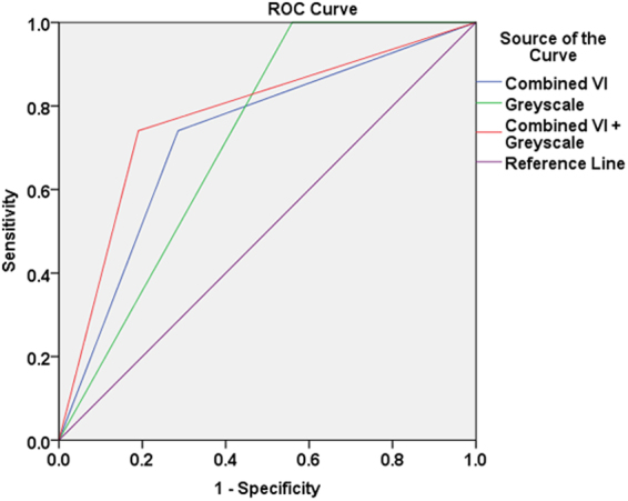
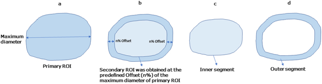
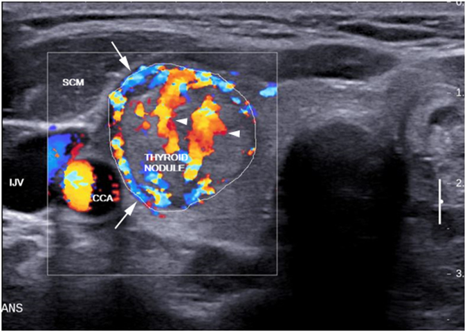
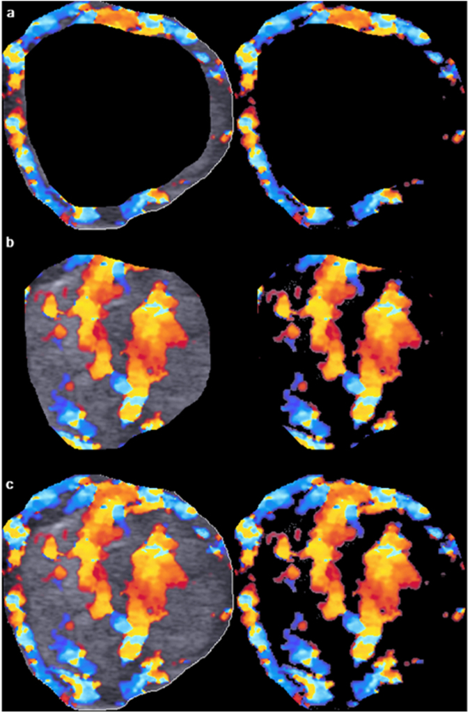
Color Doppler vascular index (VI) was assessed alone and in combination with grey-scale ultrasound (GSU) in regionally subdivided thyroid nodules in diagnosing thyroid cancer. Color Doppler sonograms of 111 thyroid nodules were evaluated by a home-developed algorithm that performed "offsetting" (algorithm for changing the area of a region of interest, ROI, without distorting the ROI's contour) and assessed peripheral, central and overall VI of thyroid nodules. Results showed that the optimum offset for dividing peripheral and central regions of nodule was 22%. At the optimum offset, the mean VI of peripheral, central, and overall regions of malignant nodules were significantly higher than those of benign nodules (26.5 ± 16.2%, 21.7 ± 19.6%, 23.8 ± 4.6% v/s 18.2 ± 16.7%, 11.9 ± 15.1% and 16.6 ± 1.8% respectively, P < 0.05). The optimum cut-off of peripheral, central, and overall VI was 19.7%, 9.1% and 20.2% respectively. When compared to GSU alone, combination of VI assessment with GSU evaluation of thyroid nodules increased the diagnostic accuracy from 58.6% to 79.3% (P < 0.05). In conclusion, a novel algorithm for regional subdivision and quantification of thyroid nodular VI in ultrasound images was established, and the optimum offset and cut-off were derived. Assessment of intranodular VI in conjunction with GSU can increase the accuracy in ultrasound diagnosis of thyroid cancer.

摘要

彩色多普勒血管指数(VI)单独评估和与灰阶超声(GSU)联合在区域性细分甲状腺结节中诊断甲状腺癌。使用自主开发的算法评估了 111 个甲状腺结节的彩色多普勒超声图像,该算法执行了“偏移”(改变感兴趣区域(ROI)面积而不改变 ROI 轮廓的算法),并评估了甲状腺结节的外周、中央和整体 VI。结果表明,用于划分结节外周和中央区域的最佳偏移量为 22%。在最佳偏移量下,恶性结节的外周、中央和整体区域的平均 VI 明显高于良性结节(26.5±16.2%、21.7±19.6%、23.8±4.6%比 18.2±16.7%、11.9±15.1%和 16.6±1.8%,P<0.05)。外周、中央和整体 VI 的最佳截断值分别为 19.7%、9.1%和 20.2%。与 GSU 单独评估相比,将 VI 评估与 GSU 评估甲状腺结节相结合,将诊断准确性从 58.6%提高到 79.3%(P<0.05)。总之,建立了一种新的超声图像甲状腺结节 VI 区域性细分和量化算法,并得出了最佳偏移量和截断值。联合 GSU 评估结节内 VI 可以提高甲状腺癌超声诊断的准确性。

https://cdn.ncbi.nlm.nih.gov/pmc/blobs/5d51/5662577/abddb6214c12/41598_2017_14432_Fig4_HTML.jpg
https://cdn.ncbi.nlm.nih.gov/pmc/blobs/5d51/5662577/2211b616f693/41598_2017_14432_Fig1_HTML.jpg
https://cdn.ncbi.nlm.nih.gov/pmc/blobs/5d51/5662577/00e4a3d8f7d3/41598_2017_14432_Fig2_HTML.jpg
https://cdn.ncbi.nlm.nih.gov/pmc/blobs/5d51/5662577/759664ad1ab9/41598_2017_14432_Fig3_HTML.jpg
https://cdn.ncbi.nlm.nih.gov/pmc/blobs/5d51/5662577/abddb6214c12/41598_2017_14432_Fig4_HTML.jpg

相似文献

1
Computer-aided assessment of regional vascularity of thyroid nodules for prediction of malignancy.计算机辅助评估甲状腺结节的区域性血管分布用于预测恶性肿瘤。
Sci Rep. 2017 Oct 30;7(1):14350. doi: 10.1038/s41598-017-14432-7.
2
Vascularity assessment of thyroid nodules by quantitative color Doppler ultrasound.通过定量彩色多普勒超声评估甲状腺结节的血管情况
Ultrasound Med Biol. 2015 May;41(5):1287-93. doi: 10.1016/j.ultrasmedbio.2015.01.001. Epub 2015 Feb 9.
3
Vascular pattern and spectral parameters of power Doppler ultrasound as predictors of malignancy risk in thyroid nodules.能量多普勒超声的血管模式和频谱参数作为甲状腺结节恶性风险的预测指标
Laryngoscope. 2008 Dec;118(12):2182-6. doi: 10.1097/MLG.0b013e3181864ae7.
4
Quantitative analysis of tumor vascularity in benign and malignant solid thyroid nodules.良性和恶性实性甲状腺结节中肿瘤血管的定量分析。
J Ultrasound Med. 2007 Jun;26(6):837-46. doi: 10.7863/jum.2007.26.6.837.
5
Power Doppler US patterns of vascularity and spectral Doppler US parameters in predicting malignancy in thyroid nodules.甲状腺结节中血管的能量多普勒超声表现及频谱多普勒超声参数在预测恶性肿瘤中的应用
Clin Radiol. 2007 Mar;62(3):245-51. doi: 10.1016/j.crad.2006.10.003.
6
Role of 99mTc-Tetrofosmin delayed scintigraphy and color Doppler sonography in characterization of solitary thyroid nodules.99mTc-替曲膦延迟闪烁扫描和彩色多普勒超声在孤立性甲状腺结节特征性诊断中的作用
Nucl Med Commun. 2007 Nov;28(11):847-51. doi: 10.1097/MNM.0b013e32827420a3.
7
Diagnosis of thyroid cancer in children: value of gray-scale and power doppler US.儿童甲状腺癌的诊断:灰阶及能量多普勒超声的价值
Radiology. 2005 May;235(2):604-13. doi: 10.1148/radiol.2352031942. Epub 2005 Mar 15.
8
Can vascularity at power Doppler US help predict thyroid malignancy?能量多普勒超声中的血流信号能否帮助预测甲状腺恶性肿瘤?
Radiology. 2010 Apr;255(1):260-9. doi: 10.1148/radiol.09091284.
9
Role of Superb Micro-Vascular Imaging in the Preoperative Evaluation of Thyroid Nodules: Comparison With Power Doppler Flow Imaging.超微血管成像在甲状腺结节术前评估中的作用:与能量多普勒血流成像的比较
J Ultrasound Med. 2017 Jul;36(7):1329-1337. doi: 10.7863/ultra.16.07004. Epub 2017 Apr 30.
10
Color Doppler sonography in cold thyroid nodules for malignancy prediction.彩色多普勒超声在甲状腺冷结节恶性预测中的应用
Acta Chir Belg. 2002 Aug;102(4):259-62. doi: 10.1080/00015458.2002.11679309.

引用本文的文献

1
Usefulness of ultrasound quantitative assessment of microvascularity in the thyroid nodule risk stratification.超声定量评估微血管在甲状腺结节风险分层中的应用价值。
J Ultrasound. 2025 Aug 14. doi: 10.1007/s40477-025-01058-z.
2
Diagnostic performance of ACR-TIRADS combined with superb microvascular imaging for differential diagnosis of mummified thyroid nodules and papillary thyroid carcinomas.美国放射学会甲状腺影像报告和数据系统(ACR-TIRADS)联合超微血管成像对木乃伊化甲状腺结节与甲状腺乳头状癌鉴别诊断的诊断效能
Endocr Connect. 2024 Feb 7;13(3). doi: 10.1530/EC-23-0388. Print 2024 Mar 1.
3
Deep learning for the diagnosis of suspicious thyroid nodules based on multimodal ultrasound images.

本文引用的文献

1
Diagnostic Accuracy of Computer-Aided Assessment of Intranodal Vascularity in Distinguishing Different Causes of Cervical Lymphadenopathy.计算机辅助评估结内血管在鉴别颈部淋巴结病不同病因中的诊断准确性
Ultrasound Med Biol. 2016 Aug;42(8):2010-6. doi: 10.1016/j.ultrasmedbio.2016.03.014. Epub 2016 Apr 27.
2
Cancer statistics, 2016.癌症统计数据,2016 年。
CA Cancer J Clin. 2016 Jan-Feb;66(1):7-30. doi: 10.3322/caac.21332. Epub 2016 Jan 7.
3
Vascularity assessment of thyroid nodules by quantitative color Doppler ultrasound.
基于多模态超声图像的深度学习用于可疑甲状腺结节的诊断
Front Oncol. 2022 Nov 8;12:1012724. doi: 10.3389/fonc.2022.1012724. eCollection 2022.
4
Diagnostic Value of AngioPLUS Microvascular Imaging in Thyroid Nodule Diagnosis Using Quantitative and Qualitative Vascularity Grading.AngioPLUS微血管成像在甲状腺结节诊断中基于定量和定性血管分级的诊断价值
Biomedicines. 2022 Jun 29;10(7):1554. doi: 10.3390/biomedicines10071554.
5
Added value of mass characteristic frequency to 2-D shear wave elastography for differentiation of benign and malignant thyroid nodules.二维剪切波弹性成像中质量特征频率对良恶性甲状腺结节的鉴别诊断价值。
Ultrasound Med Biol. 2022 Aug;48(8):1663-1671. doi: 10.1016/j.ultrasmedbio.2022.04.218. Epub 2022 Jun 4.
6
Contrast-Enhanced Ultrasound in the Differential Diagnosis and Risk Stratification of ACR TI-RADS Category 4 and 5 Thyroid Nodules With Non-Hypovascular.非低血供的ACR TI-RADS 4类和5类甲状腺结节鉴别诊断及风险分层中的超声造影
Front Oncol. 2021 May 26;11:662273. doi: 10.3389/fonc.2021.662273. eCollection 2021.
7
Comparison of Multimodal Ultrasound Imaging with Conventional Ultrasound Risk Stratification Systems in Presurgical Risk Stratification of Thyroid Nodules.多模态超声成像与传统超声风险分层系统在甲状腺结节术前风险分层中的比较
Indian J Endocrinol Metab. 2020 Nov-Dec;24(6):537-542. doi: 10.4103/ijem.IJEM_675_20. Epub 2021 Jan 12.
8
The analysis of differential diagnosis of benign and malignant thyroid nodules based on ultrasound reports.基于超声报告的甲状腺良恶性结节鉴别诊断分析
Gland Surg. 2020 Jun;9(3):653-660. doi: 10.21037/gs.2020.04.03.
9
Grayscale, subjective color Doppler, combined grayscale with subjective color Doppler in predicting thyroid carcinoma: a retrospective analysis.灰阶、彩色多普勒、灰阶与彩色多普勒联合在甲状腺癌预测中的应用:一项回顾性分析。
Braz J Otorhinolaryngol. 2022 Mar-Apr;88(2):220-227. doi: 10.1016/j.bjorl.2020.05.024. Epub 2020 Jul 9.
10
Superb microvascular imaging compared with contrast-enhanced ultrasound to assess microvessels in thyroid nodules.与对比增强超声相比,超微血管成像在评估甲状腺结节微血管方面的应用
J Med Ultrason (2001). 2020 Apr;47(2):287-297. doi: 10.1007/s10396-020-01011-z. Epub 2020 Mar 3.
通过定量彩色多普勒超声评估甲状腺结节的血管情况
Ultrasound Med Biol. 2015 May;41(5):1287-93. doi: 10.1016/j.ultrasmedbio.2015.01.001. Epub 2015 Feb 9.
4
A Comparison of the Diagnostic Accuracy and Reliability of Subjective Grading and Computer-Aided Assessment of Intranodal Vascularity in Differentiating Metastatic and Reactive Cervical Lymphadenopathy.主观分级与计算机辅助评估颈内淋巴结血管情况在鉴别转移性与反应性颈部淋巴结病中的诊断准确性及可靠性比较
Ultraschall Med. 2016 Feb;37(1):63-7. doi: 10.1055/s-0034-1384939. Epub 2014 Aug 20.
5
Quantitative analysis of dynamic power Doppler sonograms for patients with thyroid nodules.甲状腺结节患者动态能量多普勒超声的定量分析。
Ultrasound Med Biol. 2013 Sep;39(9):1543-51. doi: 10.1016/j.ultrasmedbio.2013.03.009. Epub 2013 Jun 19.
6
Utility of gray-scale ultrasound to differentiate benign from malignant thyroid nodules.灰阶超声在鉴别甲状腺良恶性结节中的应用价值。
Indian J Radiol Imaging. 2012 Jan;22(1):63-8. doi: 10.4103/0971-3026.95407.
7
Thyroid nodules: risk stratification for malignancy with ultrasound and guided biopsy.甲状腺结节:超声及引导下活检的恶性肿瘤风险分层。
Cancer Imaging. 2011 Dec 28;11(1):209-23. doi: 10.1102/1470-7330.2011.0030.
8
Tumor angiogenesis: molecular pathways and therapeutic targets.肿瘤血管生成:分子途径和治疗靶点。
Nat Med. 2011 Nov 7;17(11):1359-70. doi: 10.1038/nm.2537.
9
Principles and mechanisms of vessel normalization for cancer and other angiogenic diseases.血管正常化的原则和机制用于癌症和其他血管生成性疾病。
Nat Rev Drug Discov. 2011 Jun;10(6):417-27. doi: 10.1038/nrd3455.
10
Preoperative detection of ovarian cancer by color Doppler ultrasonography and CA 125.通过彩色多普勒超声检查和CA 125对卵巢癌进行术前检测。
Bangladesh Med Res Counc Bull. 2010 Aug;36(2):68-73. doi: 10.3329/bmrcb.v36i2.6991.