Department of Pediatrics, Immunology Division, Faculty of Medicine and Health Sciences, University of Sherbrooke, Sherbrooke, Québec J1H 5N4, Canada.
Department of Anatomy and Cell Biology, Faculty of Medicine and Health Sciences, Université de Sherbrooke, Sherbrooke, Québec J1H 5N4, Canada.
World J Gastroenterol. 2017 Sep 28;23(36):6639-6649. doi: 10.3748/wjg.v23.i36.6639.
To investigate the role of suppressor of cytokine signaling 1 (SOCS1) in regulating MET-mediated invasive potential of hepatocellular carcinoma (HCC) cells.
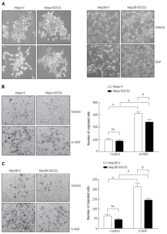
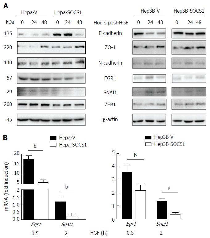
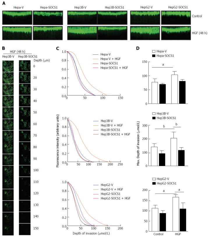
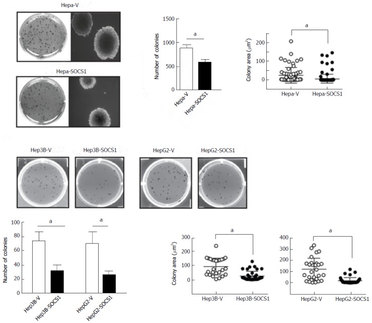
Stable derivatives of mouse (Hepa1-6) and human (hep3B, HepG2) HCC cell lines expressing SOCS1 or control vector were evaluated for their ability to migrate towards hepatocyte growth factor (HGF) in the transwell migration assay, invade extracellular matrix in response to HGF stimulation in a 3-D invasion assay by confocal microscopy, and to undergo anchorage-independent proliferation in semisolid agar. Following intravenous and intrasplenic inoculation into NOD.scid.gamma mice, the ability of Hepa cells to form othotopic tumors was evaluated. Following HGF stimulation of Hepa and Hep3B cells, expression of proteins implicated in epithelial-to-mesenchymal transition was evaluated by western blot and qRT-PCR.
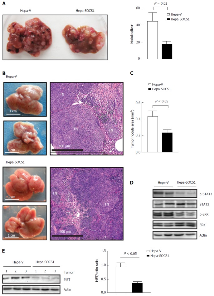
SOCS1 expression in mouse and human HCC cells inhibited HGF-induced migration through matrigel. In the 3-D invasion assay, HGF stimulation induced invasion of HCC cells across type-I collagen matrix, and SOCS1 expression significantly reduced the depth of invasion. SOCS1 expression also reduced the number and size of colonies formed by anchorage-independent growth in semisolid agar. Following intravenous inoculation, control Hepa cell formed large tumor nodules that obliterated the liver whereas the SOCS1-expressing Hepa cells formed significantly smaller nodules. Tumors formed by SOCS1-expressing cells showed reduced phosphorylation of STAT3 and ERK that was accompanied by reduced levels of MET protein expression. HGF stimulated Hepa cells expressing SOCS1 showed increased expression of E-cadherin and decreased expression of EGR1, SNAI1 and ZEB1. Comparable results were obtained with Hep3B cells. SOCS1 expressing HCC cells also showed reduced levels of EGR1 and SNAI1 transcripts.
Our findings indicate that loss of SOCS1-dependent control over epithelial-to-mesenchymal transition may contribute to MET-mediated migration, invasion and metastatic growth of HCC.
研究细胞因子信号转导抑制因子 1(SOCS1)在调节肝细胞癌(HCC)细胞中 MET 介导的侵袭潜能中的作用。
在转染实验中,用 SOCS1 或对照载体稳定转染的小鼠(Hepa1-6)和人(hep3B、HepG2)HCC 细胞系被用来检测其在 Transwell 迁移实验中向肝细胞生长因子(HGF)迁移的能力、在共聚焦显微镜下的 3-D 侵袭实验中响应 HGF 刺激侵袭细胞外基质的能力、以及在半固体琼脂中进行无锚定增殖的能力。将 Hepa 细胞静脉内和脾内接种到 NOD.scid.gamma 小鼠后,评估其形成原位肿瘤的能力。HGF 刺激 Hepa 和 Hep3B 细胞后,通过 Western blot 和 qRT-PCR 检测上皮间质转化相关蛋白的表达。
SOCS1 在小鼠和人 HCC 细胞中的表达抑制了 HGF 诱导的穿过基质胶的迁移。在 3-D 侵袭实验中,HGF 刺激诱导 HCC 细胞穿过 I 型胶原基质侵袭,SOCS1 表达显著降低了侵袭深度。SOCS1 表达还减少了半固体琼脂中无锚定生长形成的集落的数量和大小。静脉内接种后,对照 Hepa 细胞形成了大的肿瘤结节,使肝脏完全消失,而表达 SOCS1 的 Hepa 细胞形成的结节明显较小。SOCS1 表达的肿瘤显示 STAT3 和 ERK 的磷酸化减少,同时 MET 蛋白表达降低。HGF 刺激表达 SOCS1 的 Hepa 细胞显示 E-钙粘蛋白表达增加,而 EGR1、SNAI1 和 ZEB1 表达减少。在 Hep3B 细胞中也获得了类似的结果。表达 SOCS1 的 HCC 细胞也显示 EGR1 和 SNAI1 转录物水平降低。
我们的发现表明,SOCS1 依赖性对上皮间质转化的控制丧失可能导致 MET 介导的 HCC 细胞迁移、侵袭和转移生长。