UMR 7144, team EPEP, Station Biologique de Roscoff, Centre Nationnal de la Recherche Scientifique, Roscoff, France.
Université Pierre et Marie Curie, Sorbonne Universités, Roscoff, France.
Elife. 2017 Oct 31;6:e26066. doi: 10.7554/eLife.26066.
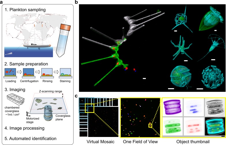
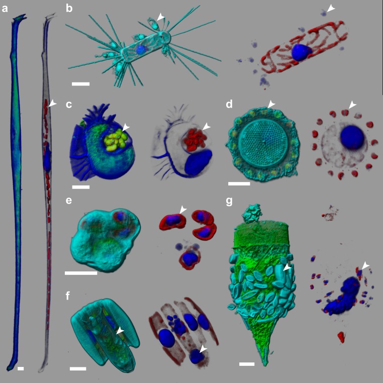
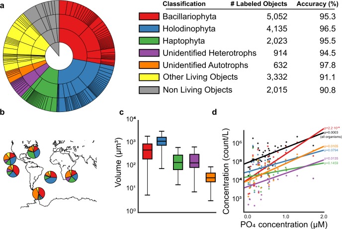
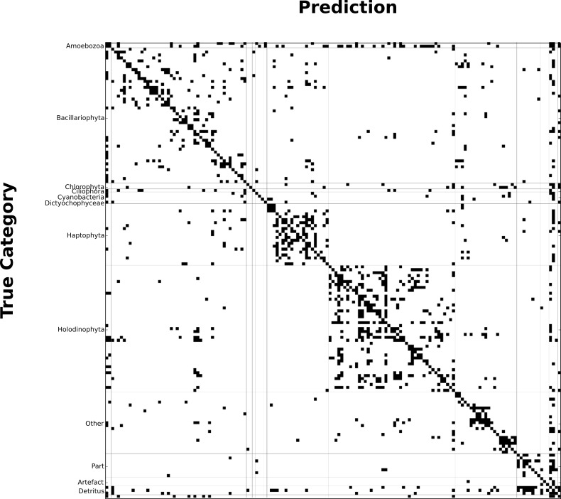
We present a 3D-fluorescence imaging and classification tool for high throughput analysis of microbial eukaryotes in environmental samples. It entails high-content feature extraction that permits accurate automated taxonomic classification and quantitative data about organism ultrastructures and interactions. Using plankton samples from the Oceans expeditions, we validate its applicability to taxonomic profiling and ecosystem analyses, and discuss its potential for future integration of eukaryotic cell biology into evolutionary and ecological studies.
我们提出了一种用于环境样本中微生物真核生物高通量分析的三维荧光成像和分类工具。它需要进行高内涵特征提取,从而实现准确的自动分类,并提供有关生物体超微结构和相互作用的定量数据。我们使用海洋考察中的浮游生物样本验证了它在分类分析和生态系统分析中的适用性,并讨论了将真核细胞生物学纳入进化和生态研究的未来整合潜力。