Rashedi Iran, Talele Nilesh, Wang Xing-Hua, Hinz Boris, Radisic Milica, Keating Armand
Institute of Biomaterials and Biomedical Engineering, University of Toronto, Toronto, Canada.
Cell Therapy Program, University Health Network, Toronto, Canada.
PLoS One. 2017 Oct 31;12(10):e0187348. doi: 10.1371/journal.pone.0187348. eCollection 2017.
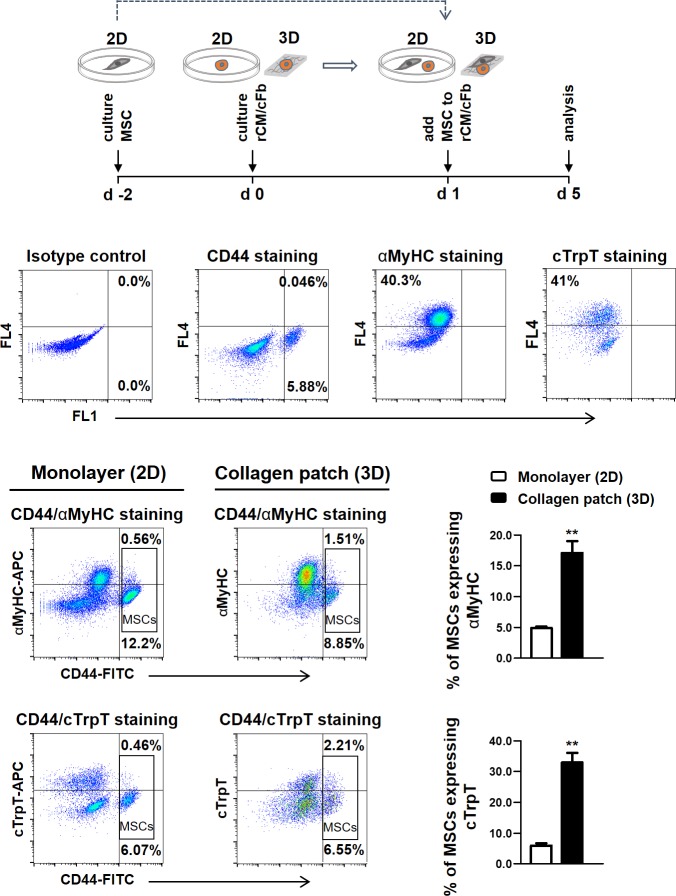
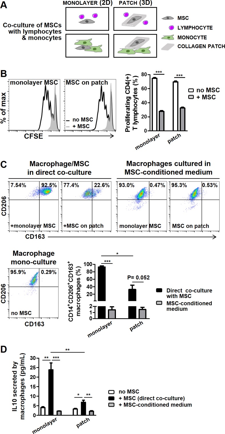
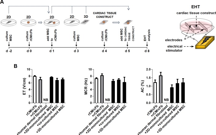
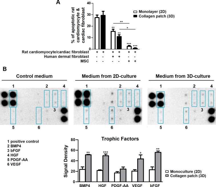
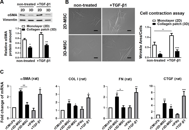
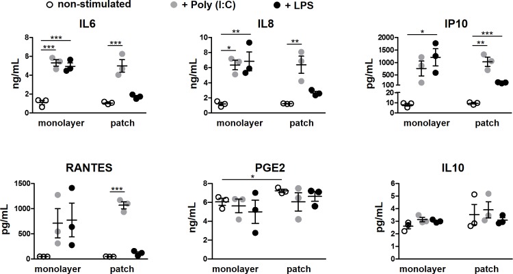
MSCs are widely applied to regenerate heart tissue in myocardial diseases but when grown in standard two-dimensional (2D) cultures exhibit limited potential for cardiac repair and develop fibrogenic features with increasing culture time. MSCs can undergo partial cardiomyogenic differentiation, which improves their cardiac repair capacity. When applied to collagen patches they may improve cardiac tissue regeneration but the mechanisms remain elusive. Here, we investigated the regenerative properties of MSCs grown in a collagen scaffold as a three-dimensional (3D) culture system, and performed functional analysis using an engineered heart tissue (EHT) model. We showed that the expression of cardiomyocyte-specific proteins by MSCs co-cultured with rat neonatal cardiomyocytes was increased in collagen patches versus conventional cultures. MSCs in 3D collagen patches were less fibrogenic, secreted more cardiotrophic factors, retained anti-apoptotic and immunomodulatory function, and responded less to TLR4 ligand lipopolysaccharide (LPS) stimulation. EHT analysis showed no effects by MSCs on cardiomyocyte function, whereas control dermal fibroblasts abrogated the beating of cardiac tissue constructs. We conclude that 3D collagen scaffold improves the cardioprotective effects of MSCs by enhancing the production of trophic factors and modifying their immune modulatory and fibrogenic phenotype. The improvement in myocardial function by MSCs after acquisition of a partial cardiac cell-like phenotype is not due to enhanced MSC contractility. A better understanding of the mechanisms of MSC-mediated tissue repair will help to further enhance the therapeutic potency of MSCs.
间充质干细胞(MSCs)被广泛应用于心肌疾病的心脏组织再生,但在标准二维(2D)培养条件下生长时,其心脏修复潜力有限,且随着培养时间的延长会出现纤维化特征。MSCs可发生部分心肌分化,从而提高其心脏修复能力。当应用于胶原蛋白贴片时,它们可能会改善心脏组织再生,但其机制仍不清楚。在此,我们研究了在胶原蛋白支架中作为三维(3D)培养系统生长的MSCs的再生特性,并使用工程心脏组织(EHT)模型进行了功能分析。我们发现,与传统培养相比,与大鼠新生心肌细胞共培养的MSCs在胶原蛋白贴片中心肌细胞特异性蛋白的表达增加。3D胶原蛋白贴片中的MSCs纤维化程度较低,分泌更多的心脏营养因子,保留抗凋亡和免疫调节功能,并且对Toll样受体4(TLR4)配体脂多糖(LPS)刺激的反应较小。EHT分析表明,MSCs对心肌细胞功能没有影响,而对照真皮成纤维细胞则消除了心脏组织构建体的跳动。我们得出结论,3D胶原蛋白支架通过增强营养因子的产生以及改变其免疫调节和纤维化表型来改善MSCs的心脏保护作用。MSCs在获得部分心肌样细胞表型后对心肌功能的改善并非由于MSCs收缩性增强。更好地理解MSCs介导的组织修复机制将有助于进一步提高MSCs的治疗效力。