Vila Natalia, Siblini Aya, Esposito Evangelina, Bravo-Filho Vasco, Zoroquiain Pablo, Aldrees Sultan, Logan Patrick, Arias Lluis, Burnier Miguel N
Henry C. Witelson Ocular Pathology Laboratory, Pathology Department, McGill University, Montreal, Canada.
Hospital Universitari de Bellvitge, Ophthalmology Department, Barcelona University, Barcelona, Spain.
BMC Ophthalmol. 2017 Nov 2;17(1):198. doi: 10.1186/s12886-017-0592-2.
Light exposure and more specifically the spectrum of blue light contribute to the oxidative stress in Age-related macular degeneration (AMD). The purpose of the study was to establish whether blue light filtering could modify proangiogenic signaling produced by retinal pigmented epithelial (RPE) cells under different conditions simulating risk factors for AMD.
Three experiments were carried out in order to expose ARPE-19 cells to white light for 48 h with and without blue light-blocking filters (BLF) in different conditions. In each experiment one group was exposed to light with no BLF protection, a second group was exposed to light with BLF protection, and a control group was not exposed to light. The ARPE-19 cells used in each experiment prior to light exposure were cultured for 24 h as follows: Experiment 1) Normoxia, Experiment 2) Hypoxia, and Experiment 3) Lutein supplemented media in normoxia. The media of all groups was harvested after light exposure for sandwich ELISA-based assays to quantify 10 pro-angiogenic cytokines.
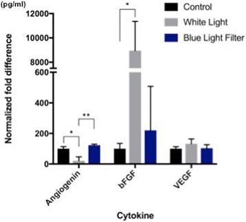
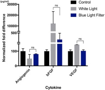
A significant decrease in angiogenin secretion levels and a significant increase in bFGF were observed following light exposure, compared to dark conditions, in both normoxia and hypoxia conditions. With the addition of a blue light-blocking filter in normoxia, a significant increase in angiogenin levels was observed. Although statistical significance was not achieved, blue light filters reduce light-induced secretion of bFGF and VEGF to near normal levels. This trend is also observed when ARPE-19 cells are grown under hypoxic conditions and when pre-treated with lutein prior to exposure to experimental conditions.
Following light exposure, there is a decrease in angiogenin secretion by ARPE-19 cells, which was abrogated with a blue light - blocking filter. Our findings support the position that blue light filtering affects the secretion of angiogenic factors by retinal pigmented epithelial cells under normoxic, hypoxic, and lutein-pretreated conditions in a similar manner.
光照,尤其是蓝光光谱,会导致年龄相关性黄斑变性(AMD)中的氧化应激。本研究的目的是确定蓝光滤过是否能改变视网膜色素上皮(RPE)细胞在模拟AMD危险因素的不同条件下产生的促血管生成信号。
进行了三项实验,以便在不同条件下,将ARPE-19细胞暴露于白光下48小时,有无蓝光阻断滤光片(BLF)。在每个实验中,一组暴露于无BLF保护的光下,第二组暴露于有BLF保护的光下,对照组不暴露于光下。每个实验中用于光照前的ARPE-19细胞培养24小时如下:实验1)常氧,实验2)缺氧,实验3)常氧条件下添加叶黄素的培养基。光照后收集所有组的培养基,用于基于夹心ELISA的检测,以定量10种促血管生成细胞因子。
与黑暗条件相比,在常氧和缺氧条件下,光照后血管生成素分泌水平显著降低,碱性成纤维细胞生长因子(bFGF)显著增加。在常氧条件下添加蓝光阻断滤光片后,观察到血管生成素水平显著增加。虽然未达到统计学显著性,但蓝光滤光片可将光诱导的bFGF和血管内皮生长因子(VEGF)分泌降低至接近正常水平。当ARPE-19细胞在缺氧条件下生长以及在暴露于实验条件之前用叶黄素预处理时,也观察到这种趋势。
光照后,ARPE-19细胞的血管生成素分泌减少,而蓝光阻断滤光片可消除这种减少。我们的研究结果支持以下观点:在常氧、缺氧和叶黄素预处理条件下,蓝光滤过以类似方式影响视网膜色素上皮细胞血管生成因子的分泌。