Tran Hai B, Jersmann Hubertus, Truong Tung Thanh, Hamon Rhys, Roscioli Eugene, Ween Miranda, Pitman Melissa R, Pitson Stuart M, Hodge Greg, Reynolds Paul N, Hodge Sandra
Lung Research Unit, Hanson Institute and Department of Thoracic Medicine, Royal Adelaide Hospital, and Department of Medicine, University of Adelaide, Adelaide, Australia.
Department of TB & Lung Diseases, Hospital 175, Hochiminh City, Vietnam.
PLoS One. 2017 Nov 7;12(11):e0179577. doi: 10.1371/journal.pone.0179577. eCollection 2017.
We have previously established a link between impaired phagocytic capacity and deregulated S1P signaling in alveolar macrophages from COPD subjects. We hypothesize that this defect may include a disruption of epithelial-macrophage crosstalk via Spns2-mediated intercellular S1P signaling.
Primary alveolar macrophages and bronchial epithelial cells from COPD subjects and controls, cell lines, and a mouse model of chronic cigarette smoke exposure were studied. Cells were exposed to 10% cigarette smoke extract, or vehicle control. Spns2 expression and subcellular localization was studied by immunofluorescence, confocal microscopy and RT-PCR. Phagocytosis was assessed by flow-cytometry. Levels of intra- and extracellular S1P were measured by S1P [3H]-labeling.
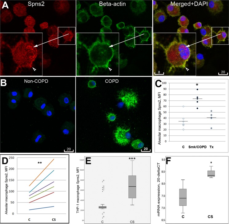
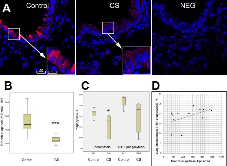
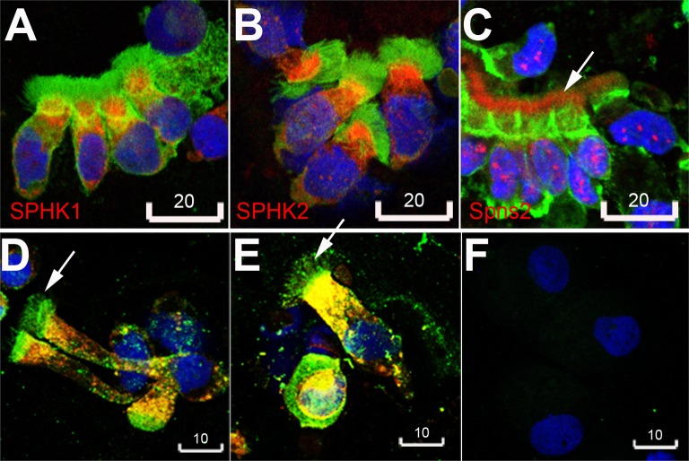
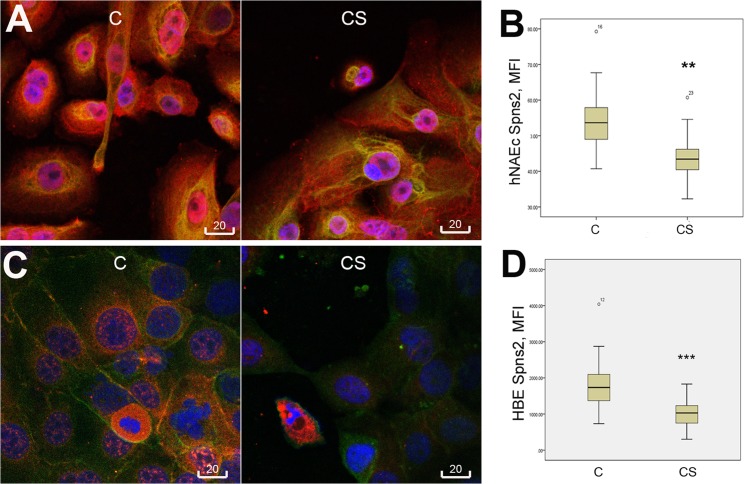
Spns2 expression was significantly increased (p<0.05) in alveolar macrophages from current-smokers/COPD patients (n = 5) compared to healthy nonsmokers (n = 8) and non-smoker lung transplant patients (n = 4). Consistent with this finding, cigarette smoke induced a significant increase in Spns2 expression in both human alveolar and THP-1 macrophages. In contrast, a remarkable Spns2 down-regulation was noted in response to cigarette smoke in 16HBE14o- cell line (p<0.001 in 3 experiments), primary nasal epithelial cells (p<0.01 in 2 experiments), and in smoke-exposed mice (p<0.001, n = 6 animals per group). Spns2 was localized to cilia in primary bronchial epithelial cells. In both macrophage and epithelial cell types, Spns2 was also found localized to cytoplasm and the nucleus, in line with a predicted bipartile Nuclear Localization Signal at the position aa282 of the human Spns2 sequence. In smoke-exposed mice, alveolar macrophage phagocytic function positively correlated with Spns2 protein expression in bronchial epithelial cells.
Our data suggest that the epithelium may be the major source for extracellular S1P in the airway and that there is a possible disruption of epithelial/macrophage cross talk via Spns2-mediated S1P signaling in COPD and in response to cigarette smoke exposure.
我们之前已经在慢性阻塞性肺疾病(COPD)患者的肺泡巨噬细胞中建立了吞噬能力受损与鞘氨醇-1-磷酸(S1P)信号失调之间的联系。我们推测,这一缺陷可能包括通过Spns2介导的细胞间S1P信号传导导致上皮细胞与巨噬细胞之间的串扰中断。
对来自COPD患者和对照的原代肺泡巨噬细胞和支气管上皮细胞、细胞系以及慢性香烟烟雾暴露小鼠模型进行了研究。将细胞暴露于10%的香烟烟雾提取物或溶剂对照中。通过免疫荧光、共聚焦显微镜和逆转录聚合酶链反应(RT-PCR)研究Spns2的表达和亚细胞定位。通过流式细胞术评估吞噬作用。通过S1P [3H]标记测量细胞内和细胞外S1P的水平。
与健康非吸烟者(n = 8)和非吸烟肺移植患者(n = 4)相比,当前吸烟者/COPD患者(n = 5)的肺泡巨噬细胞中Spns2表达显著增加(p<0.05)。与此发现一致,香烟烟雾可诱导人肺泡巨噬细胞和THP-1巨噬细胞中Spns2表达显著增加。相反,在16HBE14o-细胞系(3次实验中p<0.001)、原代鼻上皮细胞(2次实验中p<0.01)以及烟雾暴露小鼠(p<0.001,每组n = 6只动物)中,观察到香烟烟雾导致Spns2显著下调。Spns2定位于原代支气管上皮细胞的纤毛中。在巨噬细胞和上皮细胞类型中,还发现Spns2定位于细胞质和细胞核,这与人类Spns2序列第aa282位预测的双分型核定位信号一致。在烟雾暴露小鼠中,肺泡巨噬细胞吞噬功能与支气管上皮细胞中Spns2蛋白表达呈正相关。
我们的数据表明,上皮细胞可能是气道中细胞外S1P的主要来源,并且在COPD以及对香烟烟雾暴露的反应中,可能存在通过Spns2介导的S1P信号传导导致的上皮细胞/巨噬细胞串扰中断。