Sakai Chiemi, Ishida Mari, Ohba Hideo, Yamashita Hiromitsu, Uchida Hitomi, Yoshizumi Masao, Ishida Takafumi
Department of Cardiovascular Physiology and Medicine, Graduate School of Biomedical and Health Sciences, Hiroshima University, Hiroshima, Japan.
Department of Cardiovascular Medicine, Fukushima Medical University, Fukushima, Japan.
PLoS One. 2017 Nov 9;12(11):e0187934. doi: 10.1371/journal.pone.0187934. eCollection 2017.
Omega-3 fatty acids, particularly eicosapentaenoic acid (EPA) and docosahexaenoic acid (DHA), likely prevent cardiovascular disease, however their mechanisms remain unclear. Recently, the role of DNA damage in atherogenesis has been receiving considerable attention. Here, we investigated the effects of EPA and DHA on DNA damage in vascular endothelial cells to clarify their antiatherogenic mechanisms.
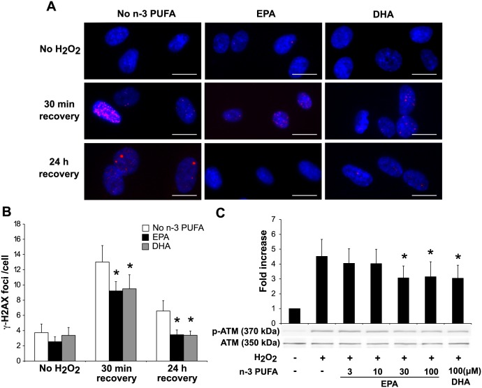
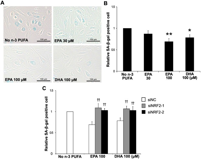
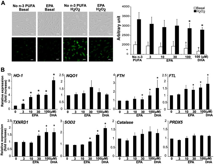
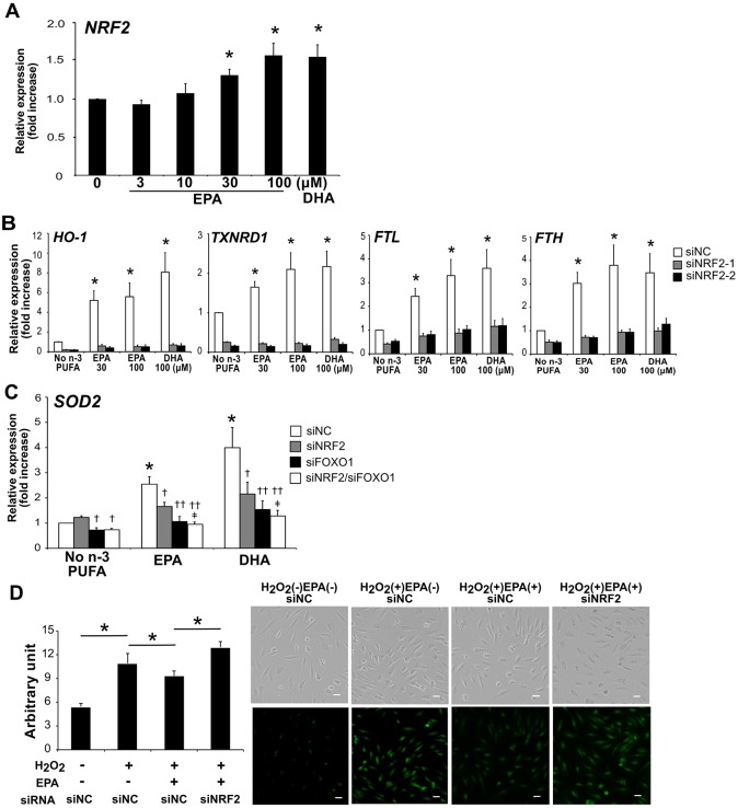
We determined the effect of EPA and DHA on H2O2-induced DNA damage response in human aortic endothelial cells. Immunofluorescence staining showed that γ-H2AX foci formation, a prominent marker of DNA damage, was significantly reduced in the cells treated with EPA and DHA (by 47% and 48%, respectively). H2O2-induced activation of ATM, a major kinase orchestrating DNA damage response, was significantly reduced with EPA and DHA treatment (by 31% and 33%, respectively). These results indicated EPA and DHA attenuated DNA damage independently of the DNA damage response. Thus the effects of EPA and DHA on a source of DNA damage were examined. EPA and DHA significantly reduced intracellular reactive oxygen species under both basal condition and H2O2 stimulation. In addition, the mRNA levels of antioxidant molecules, such as heme oxygenase-1, thioredoxin reductase 1, ferritin light chain, ferritin heavy chain and manganese superoxide dismutase, were significantly increased with EPA and DHA. Silencing nuclear factor erythroid 2-related factor 2 (NRF2) remarkably abrogated the increases in mRNA levels of antioxidant molecules and the decrease in intracellular reactive oxygen species. Furthermore, EPA and DHA significantly reduced H2O2-induced senescence-associated β-galactosidase activity in the cells (by 31% and 22%, respectively), which was revoked by NRF2 silencing.
Our results suggested that EPA and DHA attenuate oxidative stress-induced DNA damage in vascular endothelial cells through upregulation of NRF2-mediated antioxidant response. Therefore omega-3 fatty acids likely help prevent cardiovascular disease, at least in part, by their genome protective properties.
ω-3脂肪酸,尤其是二十碳五烯酸(EPA)和二十二碳六烯酸(DHA),可能预防心血管疾病,但其机制仍不清楚。最近,DNA损伤在动脉粥样硬化发生中的作用受到了相当多的关注。在此,我们研究了EPA和DHA对血管内皮细胞DNA损伤的影响,以阐明它们的抗动脉粥样硬化机制。
我们测定了EPA和DHA对人主动脉内皮细胞中过氧化氢(H2O2)诱导的DNA损伤反应的影响。免疫荧光染色显示,DNA损伤的一个显著标志物γ-H2AX焦点形成在经EPA和DHA处理的细胞中显著减少(分别减少47%和48%)。H2O2诱导的ATM(一种协调DNA损伤反应的主要激酶)激活在EPA和DHA处理后显著降低(分别降低31%和33%)。这些结果表明EPA和DHA独立于DNA损伤反应减轻了DNA损伤。因此,研究了EPA和DHA对DNA损伤来源的影响。EPA和DHA在基础条件和H2O2刺激下均显著降低细胞内活性氧。此外,EPA和DHA使抗氧化分子如血红素加氧酶-1、硫氧还蛋白还原酶1、铁蛋白轻链、铁蛋白重链和锰超氧化物歧化酶的mRNA水平显著升高。沉默核因子红细胞2相关因子2(NRF2)显著消除了抗氧化分子mRNA水平的升高和细胞内活性氧的降低。此外,EPA和DHA显著降低细胞中H2O2诱导的衰老相关β-半乳糖苷酶活性(分别降低31%和22%),而NRF2沉默可逆转这一作用。
我们的结果表明,EPA和DHA通过上调NRF2介导的抗氧化反应减轻血管内皮细胞中氧化应激诱导的DNA损伤。因此,ω-3脂肪酸可能至少部分通过其基因组保护特性有助于预防心血管疾病。