Kasalak Ömer, Glaudemans Andor W J M, Overbosch Jelle, Jutte Paul C, Kwee Thomas C
Department of Radiology, Nuclear Medicine and Molecular Imaging, University Medical Center Groningen, University of Groningen, Hanzeplein 1, PO Box 30.001, 9700 RB, Groningen, The Netherlands.
Department of Orthopedics, University Medical Center Groningen, University of Groningen, Groningen, The Netherlands.
Skeletal Radiol. 2018 Mar;47(3):363-367. doi: 10.1007/s00256-017-2807-2. Epub 2017 Nov 9.
To determine and compare the value of F-fluoro-2-deoxy-D-glucose positron emission tomography/computed tomography (FDG-PET/CT) to blind bone marrow biopsy (BMB) of the posterior iliac crest in detecting metastatic bone marrow involvement in newly diagnosed Ewing sarcoma.
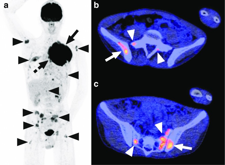
This retrospective study included 20 patients with newly diagnosed Ewing sarcoma who underwent pretreatment FDG-PET/CT and a total of 38 blind BMBs (two unilateral and 18 bilateral) of the posterior iliac crest. FDG-PET/CT scans were evaluated for bone marrow involvement, both in the posterior iliac crest and other sites, and compared to blind BMB results.
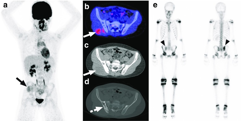
FDG-PET/CT was positive for bone marrow involvement in 7/38 posterior iliac crests, whereas BMB was positive in 5/38 posterior iliac crests. FDG-PET/CT and BMB results in the posterior iliac crest agreed in 36/38 cases (94.7%, 95% confidence interval [CI]: 82.7-98.5%). On a patient level, FDG-PET/CT was positive for bone marrow involvement in 4/20 patients, whereas BMB of the posterior iliac crest was positive in 3/20 patients. On a patient level, FDG-PET/CT and BMB results agreed in 19/20 patients (95.0%, 95% CI: 76.4-99.1%). The only discrepancies between FDG-PET/CT and BMB were observed in two BMBs of one patient. Both BMBs in this patient were negative, whereas FDG-PET/CT indicated bilateral posterior iliac crest involvement and also extensive bone marrow involvement elsewhere.
FDG-PET/CT appears to be a valuable method for metastatic bone marrow assessment in newly diagnosed Ewing sarcoma. The routine use of blind BMB of the posterior iliac crest should be reconsidered when FDG-PET/CT is available.
确定并比较F-氟-2-脱氧-D-葡萄糖正电子发射断层扫描/计算机断层扫描(FDG-PET/CT)与盲法髂后嵴骨髓活检(BMB)在检测新诊断的尤因肉瘤骨髓转移受累情况中的价值。
这项回顾性研究纳入了20例新诊断的尤因肉瘤患者,这些患者在治疗前接受了FDG-PET/CT检查,并对其进行了总共38次盲法髂后嵴骨髓活检(2次单侧活检和18次双侧活检)。对FDG-PET/CT扫描结果进行评估,以确定髂后嵴及其他部位的骨髓受累情况,并与盲法骨髓活检结果进行比较。
FDG-PET/CT显示38例髂后嵴中有7例骨髓受累呈阳性,而BMB显示38例髂后嵴中有5例呈阳性。FDG-PET/CT与BMB在髂后嵴的结果在38例中有36例一致(94.7%,95%置信区间[CI]:82.7 - 98.5%)。在患者层面,FDG-PET/CT显示20例患者中有4例骨髓受累呈阳性,而髂后嵴BMB显示20例患者中有3例呈阳性。在患者层面,FDG-PET/CT与BMB结果在20例患者中有19例一致(95.0%,95%CI:76.4 - 99.1%)。FDG-PET/CT与BMB之间的唯一差异出现在1例患者的2次BMB中。该患者的2次BMB均为阴性,而FDG-PET/CT显示双侧髂后嵴受累,且其他部位也有广泛的骨髓受累。
FDG-PET/CT似乎是评估新诊断的尤因肉瘤骨髓转移的一种有价值的方法。当有FDG-PET/CT可用时,应重新考虑常规使用盲法髂后嵴骨髓活检。