National & Regional United Engineering Lab of Tissue Engineering, Department of Orthopaedics, Southwest Hospital, the Third Military Medical University, Chongqing, China.
Department of Surgery, Fuzhou Mawei Naval Hospital, Fujian, China.
Stem Cell Res Ther. 2017 Nov 10;8(1):258. doi: 10.1186/s13287-017-0693-0.
The recruitment of a sufficient number of endogenous mesenchymal stem cells (MSCs) is the first stage of in-situ tissue regeneration. Transforming growth factor beta-3 (TGFβ3) could recruit stem or progenitor cells and endothelial cells to participate in tissue regeneration. However, the mechanism of TGFβ3 recruiting MSCs toward bone regeneration has remained obscure.
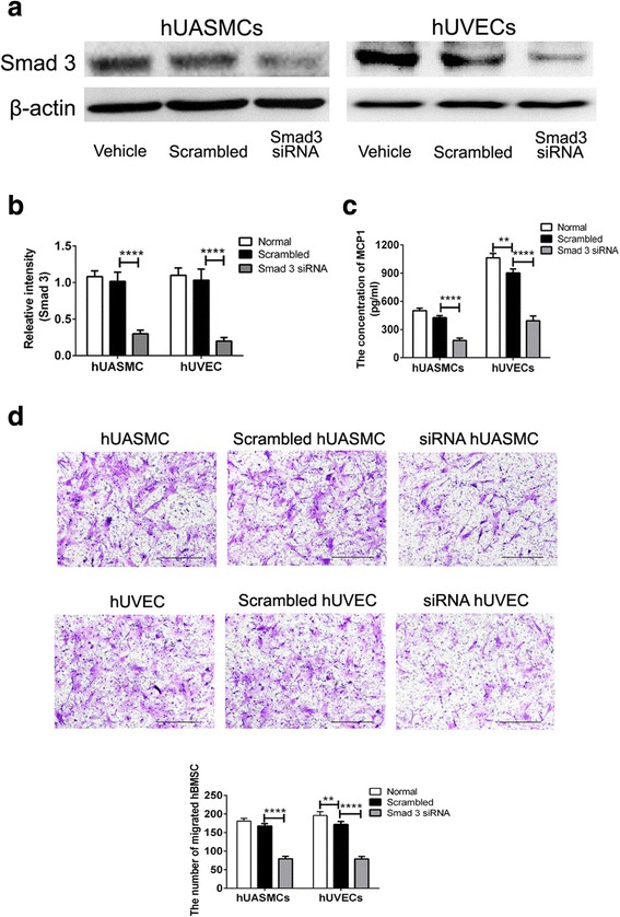
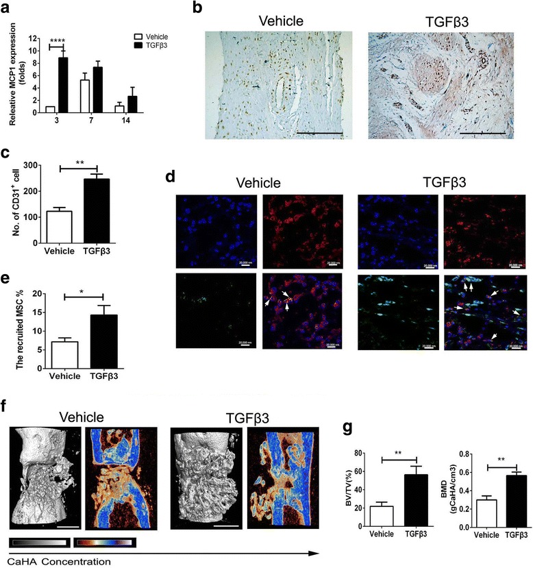
We estimated the promigratory property of TGFβ3 on human bone marrow MSCs (hBMSCs) cocultured with the vascular cells (human umbilical artery smooth muscle cells or human umbilical vein endothelial cells) or not by Transwell assay. After the addition of the inhibitor (SB431542) or Smad3 siRNA, the levels of MCP1 and SDF1 in coculture medium were tested by ELISA kit, and then the migratory signaling pathway of hBMSCs induced by TGFβ3 was investigated by western blot analysis. In vivo, a 2-mm FVB/N mouse femur defect model was used to evaluate chemokine secretion, endogenous cell homing, and bone regeneration induced by scaffolds loading 1 μg TGFβ3 through qPCR, immunofluorescent staining, immunohistochemical analysis, and Micro-CT, compared to the vehicle group.
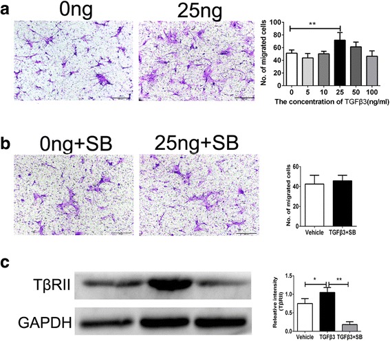
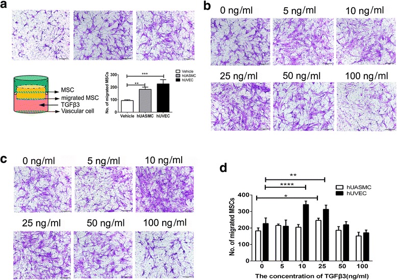
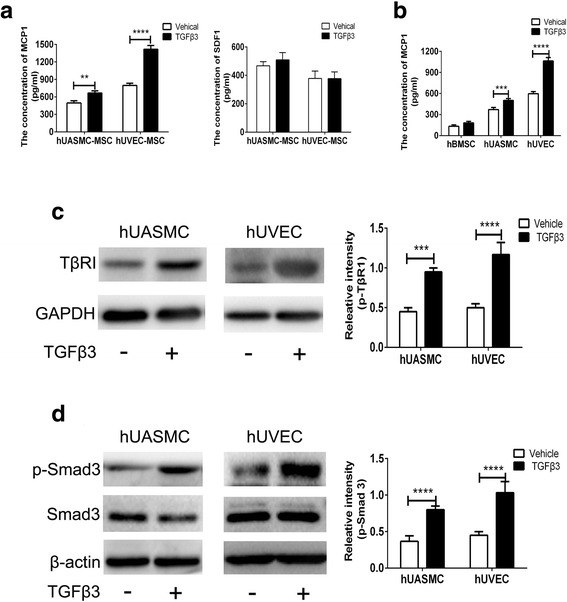
TGFβ3 (25 ng/ml) directly showed a nearly 40% increase in migrated hBMSCs via the TGFβ signaling pathway, compared to the vehicle treatment. Then, in the coculture system of hBMSCs and vascular cells, TGFβ3 further upregulated nearly 3-fold MCP1 secretion from vascular cells in a Smad3-dependent manner, to indirectly enhance nearly more than 50% of migrated hBMSCs. In vivo, TGFβ3 delivery improved MCP1 expression by nearly 7.9-fold, recruited approximately 2.0-fold CD31 vascular cells and 2.0-fold Sca-1 PDGFR-α MSCs, and achieved 2.5-fold bone volume fraction (BV/TV) and 2.0-fold bone mineral density, relative to TGFβ3-free delivery.
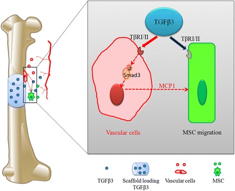
TGFβ3, as a MSC homing molecule, recruited MSCs to initiate bone formation in the direct-dependent and indirect-dependent mechanisms. This may shed light on the improvement of MSC homing in bone regeneration.
招募足够数量的内源性间充质干细胞(MSCs)是原位组织再生的第一阶段。转化生长因子β-3(TGFβ3)可以招募干细胞或祖细胞和内皮细胞参与组织再生。然而,TGFβ3 将 MSCs 募集到骨再生中的机制仍然不清楚。
我们通过 Transwell 测定法估计 TGFβ3 对与人血管细胞(人脐动脉平滑肌细胞或人脐静脉内皮细胞)共培养或不共培养的人骨髓间充质干细胞(hBMSCs)的促迁移特性。加入抑制剂(SB431542)或 Smad3 siRNA 后,通过 ELISA 试剂盒检测共培养物中 MCP1 和 SDF1 的水平,然后通过 Western blot 分析研究 TGFβ3 诱导 hBMSCs 的迁移信号通路。在体内,使用 2-mm FVB/N 小鼠股骨缺损模型通过 qPCR、免疫荧光染色、免疫组织化学分析和 Micro-CT 评估支架负载 1μg TGFβ3 诱导的趋化因子分泌、内源性细胞归巢和骨再生,与载体组相比。
与载体处理相比,TGFβ3(25ng/ml)通过 TGFβ 信号通路直接使迁移的 hBMSCs 增加近 40%。然后,在 hBMSCs 和血管细胞的共培养系统中,TGFβ3 进一步以 Smad3 依赖的方式使血管细胞中 MCP1 的分泌增加近 3 倍,从而间接增强了近 50%的迁移 hBMSCs。在体内,TGFβ3 递送使 MCP1 表达增加近 7.9 倍,募集约 2.0 倍的 CD31 血管细胞和 2.0 倍的 Sca-1 PDGFR-α MSCs,并使骨体积分数(BV/TV)增加 2.5 倍,骨密度增加 2.0 倍,与 TGFβ3 无载体递送相比。
TGFβ3 作为一种 MSC 归巢分子,通过直接依赖和间接依赖的机制募集 MSC 启动骨形成。这可能为改善骨再生中的 MSC 归巢提供思路。