Department of Neurology, Jinan Central Hospital Affiliated to Shandong University, Jinan, Shandong 250013, China.
General Medical Teaching and Research Section, Jinan Central Hospital Affiliated to Shandong University, Jinan, Shandong 250013, China.
Chin Med J (Engl). 2017 Nov 20;130(22):2726-2731. doi: 10.4103/0366-6999.218025.
Damage of the medial prefrontal cortex (mPFC) results in similar characteristics to the cognitive deficiency seen with the progress of Parkinson's disease (PD). Since the course of mPFC damage is still unclear, our study aimed to investigate the effects of melatonin (MT) on neurotoxicity in the mPFC of a rat model of PD.
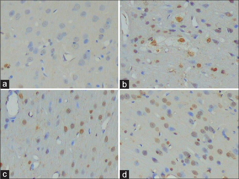
One hundred and fifty-four normal, male Wistar rats were randomly divided into the following five groups: normal + normal saline (NS), normal + 6-hydroxydopamine (6-OHDA), sham pinealectomy (PX) + 6-OHDA, PX + 6-OHDA, and MT + 6-OHDA. 6-OHDA was injected into the right substantia nigra pars compacta (SNc) and ventral tegmental area (VTA) of each group, except normal + NS, 60 days after the PX. In the MT treatment group, MT was administered immediately after the intraperitoneal injection at 4 p.m. every day, for 14 days. Neuronal apoptosis in the mPFC was examined using the TUNEL method, while the expression of tyrosine hydroxylase (TH), Bax,and Bcl-2 in this region was measured using immunohistochemistry. The concentration of malondialdehyde (MDA) in the mPFC was examined using the thiobarbituric acid method.
Rats in the normal + 6-OHDA and sham PX + 6-OHDA groups were combined into one group (Group N + 6-OHDA) since there was no significant discrepancy between the groups for all the detected parameters. Apoptosis of cells in the NS, MT + 6-OHDA, N + 6-OHDA, and PX + 6-OHDA groups was successively significantly increased (Hc = 256.25, P < 0.001). The gray value of TH (+) fibers in the NS, MT + 6-OHDA, N + 6-OHDA, and PX + 6-OHDA groups was also successively significantly increased (F = 99.33, P < 0.001). The staining intensities of Bax and Bcl-2 were as follows: Group NS +/+, Group MT + 6-OHDA ++/+, Group N + 6-OHDA ++/+, and PX + 6-OHDA +++/+. The concentrations of MDA in the NS, MT + 6-OHDA, N + 6-OHDA, and PX + 6-OHDA groups were significantly increased in sequence (Hc = 296.309, P < 0.001).
Neuronal damage of the VTA by 6-OHDA might induce VTA-mPFC nerve fibers to undergo anterograde nerve damage, in turn inducing transneuronal damage of the mPFC. PX significantly exacerbated the neurotoxicity in the mPFC, which was induced by the neuronal injury of the VTA. However, MT replacement therapy significantly alleviated the neurotoxicity in the mPFC.
内侧前额叶皮层(mPFC)的损伤导致类似于帕金森病(PD)进展过程中出现的认知缺陷。由于 mPFC 损伤的过程尚不清楚,我们的研究旨在探讨褪黑素(MT)对 PD 大鼠模型 mPFC 神经毒性的影响。
154 只正常雄性 Wistar 大鼠随机分为以下五组:正常+生理盐水(NS)、正常+6-羟多巴胺(6-OHDA)、假去松果体(PX)+6-OHDA、PX+6-OHDA 和 MT+6-OHDA。每组除 NS+正常外,于 60 天后均将 6-OHDA 注入右侧黑质致密部(SNc)和腹侧被盖区(VTA)。MT 治疗组于每日下午 4 点腹腔注射后立即给予 MT,连续 14 天。用 TUNEL 法检测 mPFC 神经元凋亡,免疫组化法检测该区域酪氨酸羟化酶(TH)、Bax 和 Bcl-2 的表达。用硫代巴比妥酸法检测 mPFC 丙二醛(MDA)浓度。
由于各组检测参数无显著差异,故将正常+6-OHDA 组和假去松果体+6-OHDA 组合并为一组(组 N+6-OHDA)。NS、MT+6-OHDA、N+6-OHDA 和 PX+6-OHDA 组细胞凋亡依次明显增加(Hc=256.25,P<0.001)。NS、MT+6-OHDA、N+6-OHDA 和 PX+6-OHDA 组 TH(+)纤维灰度值也依次明显升高(F=99.33,P<0.001)。Bax 和 Bcl-2 的染色强度依次为:组 NS++/+,组 MT+6-OHDA+++/+,组 N+6-OHDA+++/+,组 PX+6-OHDA+++/+。NS、MT+6-OHDA、N+6-OHDA 和 PX+6-OHDA 组 MDA 浓度依次明显升高(Hc=296.309,P<0.001)。
6-OHDA 对 VTA 的神经元损伤可能导致 VTA-mPFC 神经纤维发生逆行性神经损伤,进而导致 mPFC 的跨神经元损伤。去松果体显著加重了由 VTA 神经元损伤引起的 mPFC 神经毒性,而 MT 替代治疗显著减轻了 mPFC 的神经毒性。