Bakopoulos Anargyros, Kapelouzou Alkistis, Tsilimigras Diamantis I, Katsimpoulas Michalis, Schizas Dimitrios, Aravanis Chrysostomos, Balafas Evaggelos, Mavroidis Manolis, Pavlakis Kitty, Machairas Anastasios, Liakakos Theodore
Third Department of Surgery, Attikon General Hospital, School of Medicine, National and Kapodistrian University of Athens, Athens, Greece.
Center for Clinical, Experimental Surgery and Translational Research, Biomedical Research Foundation of the Academy of Athens, Athens, Greece.
PLoS One. 2017 Nov 14;12(11):e0188050. doi: 10.1371/journal.pone.0188050. eCollection 2017.
Sepsis is a condition characterized by high mortality rates and often accompanied by multiple-organ dysfunction. During sepsis, respiratory system may be affected and possibly result in acute respiratory distress syndrome (ARDS). Toll-like receptors (TLRs), as a first line defense against invading pathogens, seem to be highly expressed in septic states. Therefore, expression of TLRs in the lungs of a sepsis animal model could indicate the involvement of the respiratory system and appear as a severity index of the clinical course.
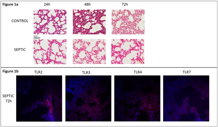
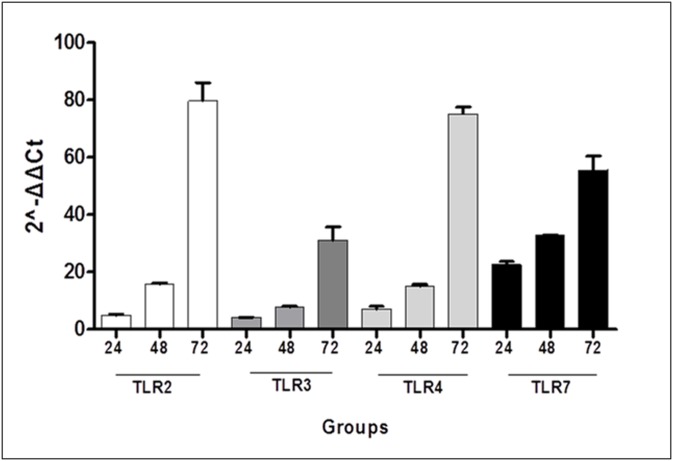
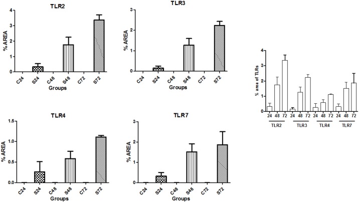
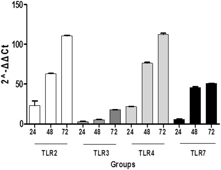
A total of 72 C57BL/6J mice, aged 12-14 weeks, were studied. The animals were divided into 3 sepsis (S) groups (24h, 48h and 72h) and 3 control (C) groups (24h, 48h and 72h), each consisting of 12 mice. The S-groups were subjected to cecal ligation and puncture (CLP) while the C-groups had a sham operation performed. Blood samples were drawn from all groups. Total blood count analysis was performed along with the measurement of certain biochemical markers. Additionally, lung tissues were harvested and the expression of TLRs, namely TLR 2, TLR 3, TLR 4 and TLR 7 were evaluated by means of immunofluorescence (IF) and qRT-PCR (quantitative-Polymerase Chain Reaction). Statistical analysis was performed by using one-way ANOVA followed by student t-test. Results were considered statistically significant when p<0.05.
WBCs and lymphocytes were decreased in all S-groups compared to the corresponding C-groups (p<0.05), while RBCs showed a gradual decline in S-groups with the lowest levels appearing in the S72 group. Only, monocytes were higher in S-groups, especially between S48-C48 (p<0.05) and S72-C72 (p<0.05). Creatinine, IL-10 and IL-6 levels were significantly increased in the S-groups compared to the corresponding C-groups (S24 vs C24, S48 vs C48 and S72 vs C72, p<0.05). IF showed that expression of TLRs 2, 3, 4 and 7 was increased in all S-groups compared to the time-adjusted C-groups (p<0.05). Similarly, qRT-PCR revealed that expression of all TLRs was higher in all S-groups compared to their respective C-groups in both lungs and intestine (p<0.05). Comparing lung and intestinal tissues from S-groups, TLRs 2 and 4 were found increased in the lung at 24, 48 and 72 hours (p<0.05), whereas TLR 3 was higher in the intestine at all time points examined (p<0.05). Finally, TLR 7 levels were significantly higher in the intestinal tissues at 24 hours (p<0.0001), while lungs predominated at 48 hours (p<0.0001).
TLRs seem to be highly expressed in the lungs of septic mice, therefore suggesting a potential role in the pathogenesis of ARDS during sepsis. While more studies need to be conducted in order to completely understand the underlying mechanisms, TLRs may represent a promising target for establishing novel therapeutic strategies in the treatment of sepsis.
脓毒症是一种死亡率高且常伴有多器官功能障碍的病症。在脓毒症期间,呼吸系统可能受到影响,并可能导致急性呼吸窘迫综合征(ARDS)。Toll样受体(TLRs)作为抵御入侵病原体的第一道防线,在脓毒症状态下似乎高度表达。因此,脓毒症动物模型肺中TLRs的表达可能表明呼吸系统的参与情况,并可作为临床病程严重程度的指标。
共研究72只12 - 14周龄的C57BL/6J小鼠。将动物分为3个脓毒症(S)组(24小时、48小时和72小时)和3个对照组(C)组(24小时、48小时和72小时),每组12只小鼠。S组进行盲肠结扎和穿刺(CLP),而C组进行假手术。从所有组采集血样。进行全血细胞计数分析以及某些生化标志物的测量。此外,采集肺组织,并通过免疫荧光(IF)和定量聚合酶链反应(qRT-PCR)评估TLRs(即TLR 2、TLR 3、TLR 4和TLR 7)的表达。采用单因素方差分析,随后进行学生t检验进行统计分析。当p < 0.05时,结果被认为具有统计学意义。
与相应的C组相比,所有S组的白细胞和淋巴细胞均减少(p < 0.05),而红细胞在S组中呈逐渐下降趋势,最低水平出现在S72组。仅单核细胞在S组中较高,尤其是在S48 - C48(p < 0.05)和S72 - C72(p < 0.05)之间。与相应的C组相比,S组的肌酐、IL - 10和IL - 6水平显著升高(S24与C24、S48与C48以及S72与C72,p < 0.05)。免疫荧光显示,与时间匹配的C组相比,所有S组中TLR 2、3、4和7的表达均增加(p < 0.05)。同样,qRT-PCR显示,与各自的C组相比,所有S组在肺和肠道中所有TLRs的表达均更高(p < 0.05)。比较S组的肺和肠道组织,发现TLR 2和4在24、48和72小时时肺中增加(p < 0.05),而TLR 3在所有检查时间点肠道中均较高(p < 0.05)。最后,TLR 7水平在24小时时肠道组织中显著更高(p < 0.0001),而在48小时时肺中占主导(p < 0.0001)。
TLRs似乎在脓毒症小鼠的肺中高度表达,因此提示其在脓毒症期间ARDS发病机制中可能发挥作用。虽然需要进行更多研究以完全了解潜在机制,但TLRs可能是建立脓毒症治疗新策略的有希望的靶点。