Department of Pathology, University of Pittsburgh, Pittsburgh, PA, USA.
McGowan Institute for Regenerative Medicine, University of Pittsburgh, Pittsburgh, PA, USA.
Mol Cancer. 2017 Nov 14;16(1):172. doi: 10.1186/s12943-017-0740-6.
Our understanding of the multiple roles exosomes play during tumor progression is still very poor and the contribution of the normal tissue derived exosomes in distant seeding and tumor outgrowth has also not been widely appreciated.
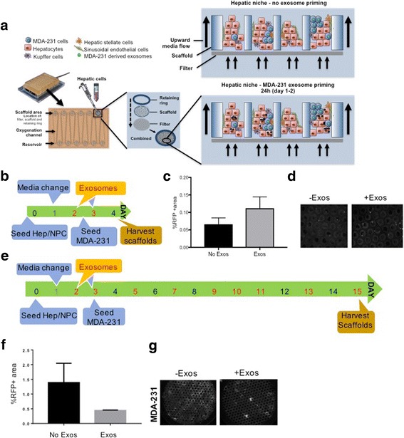
Using our all-human liver microphysiological system (MPS) platform as a model to closely recapitulate the early metastatic events, we isolated exosomes from both tumor cells and liver microenvironment.
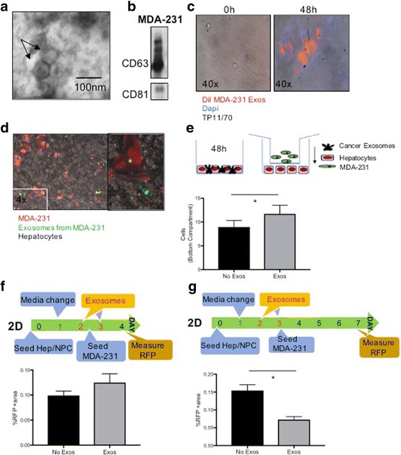
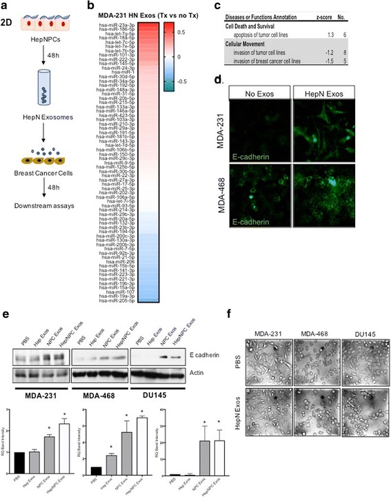
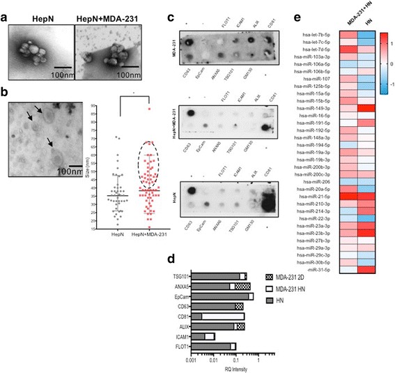
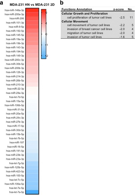
We observed that while priming of the hepatic niche (HepN) with MDA-231 breast cancer derived exosomes facilitated seeding of the cancer cells in the liver, subsequent tumor outgrowth was diminished; this was consistent with increased entry into dormancy. We found that hepatic niche (HepN) derived exosomes contribute significantly to the exosome pool and are distinguished from cancer derived exosomes based on their size, protein and miRNA content. By Ingenuity Pathway Analysis (IPA) of the miRNA content of the HepN, MDA-231/HepN and MDA-231 cells we showed that the HepN derived exosomes affect the breast cancer cells by suppressing pathways involved in cancer cell proliferation and invasion. More importantly exposure of MDA-231 and MDA-468 cells to purified normal HepN derived exosomes, induced changes in the cells consistent with a Mesenchymal to Epithelial reverting Transition (MErT). miRNA arrays performed on MDA-231 treated with Hum Hep/NPC derived exosomes showed significant changes in the levels of a select number of miRNAs involved in epithelial cell differentiation and miRNAs, such as miR186, miR23a and miR205, from our top and bottom bins have previously been reported to regulate E-cadherin transcription and MErT induction in various cancer types. Consistently HepN derived exosome treatment of breast and prostate cancer lines lead to a transient induction of E-cadherin and ZO-1 at the protein level and a more epithelial-like morphology of the cells.
Collectively our data revealed a novel mechanism of regulation of the metastatic cascade, showing a well-orchestrated, timely controlled crosstalk between the cancer cells and the HepN and implicating for the first time the normal tissue/HepN derived exosomes in enabling seeding and entry into dormancy of the cancer cells at the metastatic site.
我们对细胞外囊泡在肿瘤进展中所扮演的多种角色的理解还非常有限,而且正常组织来源的细胞外囊泡在远处播种和肿瘤生长中的作用也尚未得到广泛认可。
我们使用全人源肝脏微生理系统(MPS)平台作为模型来密切重现早期转移事件,从肿瘤细胞和肝脏微环境中分离细胞外囊泡。
我们观察到,用 MDA-231 乳腺癌衍生的细胞外囊泡对肝基质(HepN)进行预刺激,促进了癌细胞在肝脏中的播种,但随后肿瘤的生长减少;这与进入休眠相一致。我们发现,肝基质(HepN)衍生的细胞外囊泡对细胞外囊泡池的贡献很大,并且可以基于其大小、蛋白质和 miRNA 含量与癌症衍生的细胞外囊泡区分开来。通过 IPA 分析 HepN、MDA-231/HepN 和 MDA-231 细胞的 HepN 衍生细胞外囊泡的 miRNA 内容,我们表明 HepN 衍生的细胞外囊泡通过抑制参与癌细胞增殖和侵袭的途径来影响乳腺癌细胞。更重要的是,将 MDA-231 和 MDA-468 细胞暴露于纯化的正常 HepN 衍生的细胞外囊泡中,诱导细胞发生与间充质到上皮转化(MErT)相一致的变化。用 Hum Hep/NPC 衍生的细胞外囊泡处理 MDA-231 后进行 miRNA 阵列分析,显示了参与上皮细胞分化的一些 miRNA 水平的显著变化,并且我们的 top 和 bottom bins 中的 miRNA,如 miR186、miR23a 和 miR205,以前曾报道过可调节各种癌症类型的 E-钙粘蛋白转录和 MErT 诱导。一致地,HepN 衍生的细胞外囊泡处理乳腺癌和前列腺癌细胞系导致细胞内 E-钙粘蛋白和 ZO-1 的蛋白水平的短暂诱导,以及细胞更上皮样的形态。
总的来说,我们的数据揭示了一种调节转移级联的新机制,显示了癌细胞与 HepN 之间协调一致、适时的相互作用,并首次表明正常组织/HepN 衍生的细胞外囊泡在允许癌细胞在转移部位播种和进入休眠状态方面发挥作用。