Cheytan Tasneem, Schneider Martin, Würth Roberto, Schwerd-Kleine Paul, Gutjahr Ewgenija, Thewes Verena, Michel Laura L, Weber Rebecca, Vorberg Tim, Lohr Sabrina, Nitschke Katja, Neßling Michelle, Lichter Peter, Schneeweiss Andreas, Richter Karsten, Helm Dominic, Sprick Martin, Trumpp Andreas
Division of Stem Cells and Cancer, German Cancer Research Center (DKFZ) and DKFZ-ZMBH Alliance, Heidelberg, Germany.
Heidelberg Institute for Stem Cell Technology and Experimental Medicine (HI-STEM gGmbH), Heidelberg, Germany.
Mol Cancer. 2025 Mar 8;24(1):72. doi: 10.1186/s12943-025-02235-8.
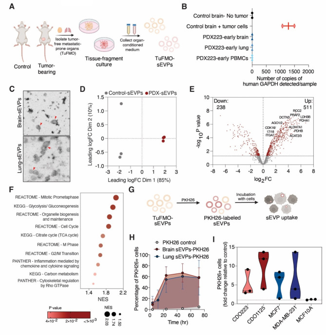
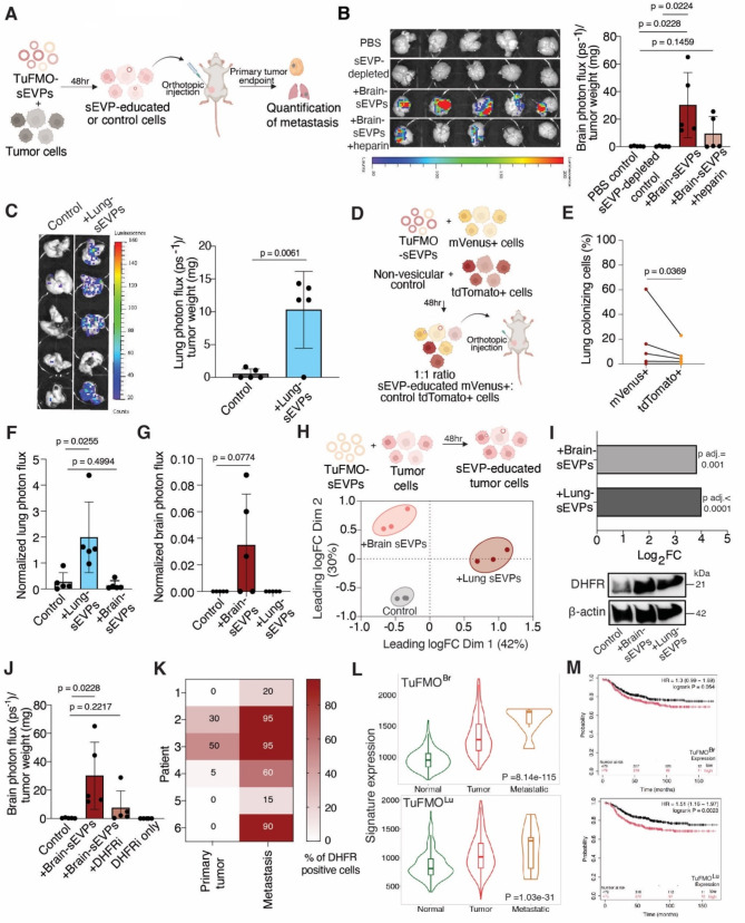
Metastatic breast cancer remains largely incurable, partly due to our incomplete understanding of its intricate underlying mechanisms. Notably, intercellular communication mediated by small extracellular vesicles and particles (sEVPs) has emerged as a key feature of metastasis. While tumor-derived sEVPs have been extensively studied and are known to be pro-metastatic, the role of sEVPs from metastasis-prone normal tissue sites remains primarily undefined. Here, we characterized and studied the function of sEVPs secreted from tumor-free pre-metastatic organs (TuFMO-sEVPs) such as the brain and lungs in both immunocompetent and patient-derived xenograft models. TuFMO-sEVPs from the brain of mammary tumor-bearing mice were found to have a distinct protein content as compared to brain-sEVPs from tumor-free mice, suggesting that the primary tumor can systemically influence the cargo of TuFMO-sEVPs. Importantly, mice orthotopically injected with breast cancer cells which had been educated with either brain or lung TuFMO-sEVPs prior to transplantation showed significantly increased metastasis to the respective organ. We further demonstrated that TuFMO-sEVPs induced the expression of the enzyme dihydrofolate reductase (DHFR) upon uptake by breast cancer cells, leading to their enhanced metastatic capacity. Organ-specific signatures generated from TuFMO-sEVP educated tumor cells were found to be increased in metastatic samples from breast cancer patients as compared to the primary tumor or normal tissue samples and these signatures also significantly correlated with poorer patient outcome. Collectively, our data reveals a novel facet of the metastatic cascade, implicating a role for TuFMO-sEVPs in directing metastasis and providing a potential therapeutic strategy for targeting this process.
转移性乳腺癌在很大程度上仍然无法治愈,部分原因是我们对其复杂的潜在机制了解不全面。值得注意的是,由小细胞外囊泡和颗粒(sEVPs)介导的细胞间通讯已成为转移的一个关键特征。虽然肿瘤来源的sEVPs已得到广泛研究且已知具有促转移作用,但来自易发生转移的正常组织部位的sEVPs的作用仍主要未明确。在此,我们在免疫健全和患者来源的异种移植模型中,对来自无肿瘤的转移前器官(如脑和肺)分泌的sEVPs(TuFMO - sEVPs)的功能进行了表征和研究。发现荷乳腺肿瘤小鼠脑内的TuFMO - sEVPs与无肿瘤小鼠脑内的sEVPs相比,具有独特的蛋白质含量,这表明原发性肿瘤可系统性地影响TuFMO - sEVPs的货物成分。重要的是,在移植前用脑或肺TuFMO - sEVPs预处理的乳腺癌细胞原位注射的小鼠,向相应器官的转移显著增加。我们进一步证明,TuFMO - sEVPs被乳腺癌细胞摄取后可诱导二氢叶酸还原酶(DHFR)的表达,从而导致其转移能力增强。与原发性肿瘤或正常组织样本相比,在乳腺癌患者的转移样本中,由TuFMO - sEVPs预处理的肿瘤细胞产生的器官特异性特征增加,并且这些特征也与患者较差的预后显著相关。总体而言,我们的数据揭示了转移级联反应的一个新方面,表明TuFMO - sEVPs在指导转移中起作用,并为靶向这一过程提供了潜在的治疗策略。