Larsen C P, Boils C L, Cossey L N, Sharma S G, Walker P D
Arkana Laboratories, Little Rock, Arkansas, USA.
Kidney Int Rep. 2016 Aug 24;1(4):299-305. doi: 10.1016/j.ekir.2016.08.012. eCollection 2016 Nov.
Ig deposits identified on renal biopsy samples by paraffin immunofluorescence that show negative staining by routine immunofluorescence on frozen tissue have become known as "masked" deposits. Membranous-like glomerulopathy with masked IgG kappa (κ) deposits is a recently recognized pattern of immune complex deposition characterized by masked deposits that show IgG κ restriction and are subepithelial and mesangial by electron microscopy. Based on the frequent presence of C3-only staining by routine immunofluorescence microscopy (IF), these cases could be misdiagnosed as C3 glomerulonephritis in the absence of paraffin immunofluorescence evaluation.
The clinicopathologic details of all cases of membranous-like glomerulopathy with masked IgG κ deposits diagnosed in our laboratory were included, beginning with the initial recognition of this entity in 2011 through the end of 2015. Inclusion was based on renal biopsy sample morphologic features including glomerular deposits that stain for IgG κ and have a staining intensity that is significantly brighter by paraffin IF than by routine IF on frozen tissue.
This pattern of immune complex deposition has been seen in 41 patients in our laboratory over a 5-year period. The patients with these biopsy findings are most commonly young female individuals with a mean age of 27.5 years, with 88% being less than 40 years. All patients had proteinuria with a mean 24-hour urine protein of 3.5 g (range 0.5-12.8 years) and 35% with nephrotic-range proteinuria. Hematuria was present in 88% of patients, and 29% had elevated serum creatinine at presentation. Autoimmune serologic tests were positive in 55% of patients, with a weakly positive antinuclear antibody being most common. Despite this, only 1 patient (2%) fulfilled the diagnostic criteria for systemic lupus erythematosus. The outcome data were mixed, as some patients showed spontaneous remission and mild disease whereas others progressed to end-stage renal disease. There was no apparent correlation between the treatment used and outcome in this retrospective analysis. One patient underwent transplantation and developed biopsy-proven recurrence of disease in the graft 42 months posttransplantation. The etiology of this entity remains unknown.
We provide an expanded case series detailing the clinicopathologic findings of patients with membranous-like glomerulopathy with masked IgG κ deposits. Patients are most commonly young female individuals <40 years of age and commonly have positive autoimmune serologic studies such as antinuclear antibody, although few carry a diagnosis of any well-defined autoimmune disease such as lupus. The outcome data were mixed, as some patients showed spontaneous remission and mild disease whereas others progressed to ESRD. There was no apparent correlation between the treatment used and outcome in this retrospective analysis.
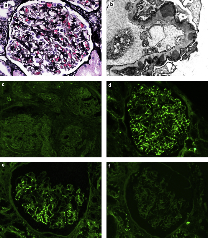
在肾活检样本中通过石蜡免疫荧光鉴定出的免疫球蛋白(Ig)沉积物,在冰冻组织上的常规免疫荧光显示为阴性染色,这种沉积物被称为“隐匿性”沉积物。伴有隐匿性IgG κ沉积物的膜性肾病样肾小球病是一种最近才被认识的免疫复合物沉积模式,其特征为隐匿性沉积物显示IgG κ受限,且电子显微镜下位于上皮下和系膜区。基于常规免疫荧光显微镜检查(IF)中仅C3染色常见,在缺乏石蜡免疫荧光评估的情况下,这些病例可能被误诊为C3肾小球肾炎。
纳入了我们实验室诊断的所有伴有隐匿性IgG κ沉积物的膜性肾病样肾小球病病例的临床病理细节,时间从2011年首次认识到该实体开始至2015年底。纳入标准基于肾活检样本的形态学特征,包括对IgG κ染色的肾小球沉积物,且其染色强度通过石蜡免疫荧光比冰冻组织上的常规免疫荧光明显更强。
在我们实验室5年期间共发现41例这种免疫复合物沉积模式的病例。有这些活检结果的患者最常见于年轻女性,平均年龄27.5岁,88%的患者年龄小于40岁。所有患者均有蛋白尿,24小时尿蛋白平均为3.5 g(范围0.5 - 12.8 g),35%的患者有肾病范围的蛋白尿。88%的患者有血尿,29%的患者就诊时血清肌酐升高。55%的患者自身免疫血清学检查呈阳性,最常见的是抗核抗体弱阳性。尽管如此,只有1例患者(2%)符合系统性红斑狼疮的诊断标准。结果数据不一,一些患者出现自发缓解且病情较轻,而另一些患者进展至终末期肾病。在这项回顾性分析中,所用治疗方法与结果之间没有明显相关性。1例患者接受了移植,移植后42个月移植肾活检证实疾病复发。该实体的病因仍然不明。
我们提供了一个扩展的病例系列,详细描述了伴有隐匿性IgG κ沉积物的膜性肾病样肾小球病患者的临床病理表现。患者最常见于年龄小于40岁的年轻女性,通常自身免疫血清学检查呈阳性,如抗核抗体,尽管很少被诊断为任何明确的自身免疫性疾病,如狼疮。结果数据不一,一些患者出现自发缓解且病情较轻,而另一些患者进展至终末期肾病。在这项回顾性分析中,所用治疗方法与结果之间没有明显相关性。