Xu Liang, Hu Yan-Xi, Li Yan-Cheng, Liu Yu-Feng, Zhang Li, Ai Hai-Xin, Liu Hong-Sheng
College of Pharmacy, Liaoning University, Shenyang, 110036, People's Republic of China.
Natural Products Pharmaceutical Engineering Technology Research Center of Liaoning Province, Shenyang, 110036, People's Republic of China.
Chem Cent J. 2017 Nov 17;11(1):116. doi: 10.1186/s13065-017-0348-3.
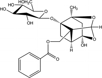
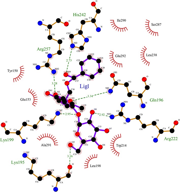
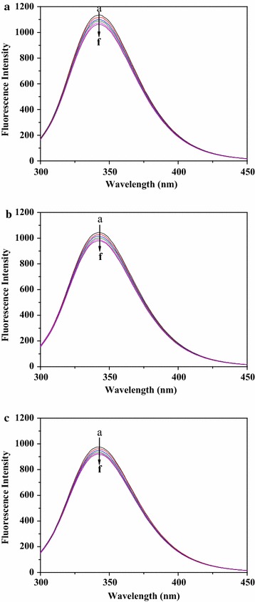
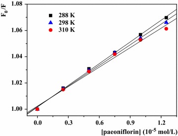
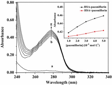
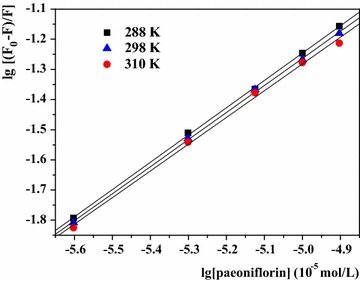
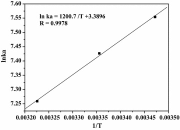
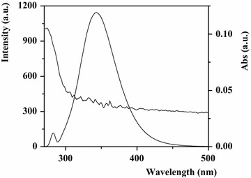
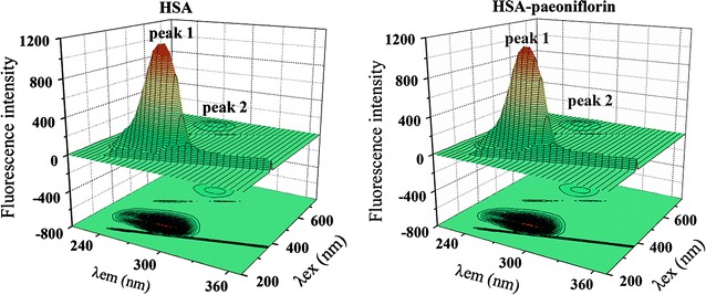
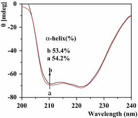
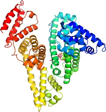
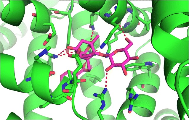
The interaction of paeoniflorin with human serum albumin (HSA) was investigated using fluorescence, UV-vis absorption, circular dichroism (CD) spectra and molecular docking techniques under simulative physiological conditions. The results clarified that the fluorescence quenching of HSA by paeoniflorin was a static quenching process and energy transfer as a result of a newly formed complex (1:1). Paeoniflorin spontaneously bound to HSA in site I (subdomain IIA), which was primarily driven by hydrophobic forces and hydrogen bonds (ΔH° = - 9.98 kJ mol, ΔS° = 28.18 J mol K). The binding constant was calculated to be 1.909 × 10 L mol at 288 K and it decreased with the increase of the temperature. The binding distance was estimated to be 1.74 nm at 288 K, showing the occurrence of fluorescence energy transfer. The results of CD and three-dimensional fluorescence spectra showed that paeoniflorin induced the conformational changes of HSA. Meanwhile, the study of molecular docking also indicated that paeoniflorin could bind to the site I of HSA mainly by hydrophobic and hydrogen bond interactions.
在模拟生理条件下,采用荧光、紫外可见吸收、圆二色光谱(CD)和分子对接技术研究了芍药苷与人血清白蛋白(HSA)的相互作用。结果表明,芍药苷对HSA的荧光猝灭是一个静态猝灭过程,由于新形成的复合物(1:1)导致能量转移。芍药苷自发地结合到HSA的位点I(亚结构域IIA),这主要是由疏水作用力和氢键驱动的(ΔH° = - 9.98 kJ/mol,ΔS° = 28.18 J/mol·K)。在288 K时计算得到结合常数为1.909×10⁴ L/mol,并且随着温度的升高而降低。在288 K时估计结合距离为1.74 nm,表明发生了荧光能量转移。CD光谱和三维荧光光谱结果表明芍药苷诱导了HSA的构象变化。同时,分子对接研究也表明芍药苷可以主要通过疏水和氢键相互作用结合到HSA的位点I。