Ahmed Mona, Cerroni Barbara, Razuvaev Anton, Härmark Johan, Paradossi Gaio, Caidahl Kenneth, Gustafsson Björn
Department of Molecular Medicine and Surgery, Karolinska Institutet, Stockholm, Sweden.
Department of Chemical Sciences and Technologies, University of Rome Tor Vergata, Rome, Italy.
Cell Mol Bioeng. 2017;10(6):537-548. doi: 10.1007/s12195-017-0504-9. Epub 2017 Aug 10.
Both diagnostic ultrasound (US) and magnetic resonance imaging (MRI) accuracy can be improved by using contrast enhancement. For US gas-filled microbubbles (MBs) or silica nanoparticles (SiNPs), and for MRI superparamagnetic or paramagnetic agents, contribute to this. However, interactions of MBs with the vascular wall and cells are not fully known for all contrast media.
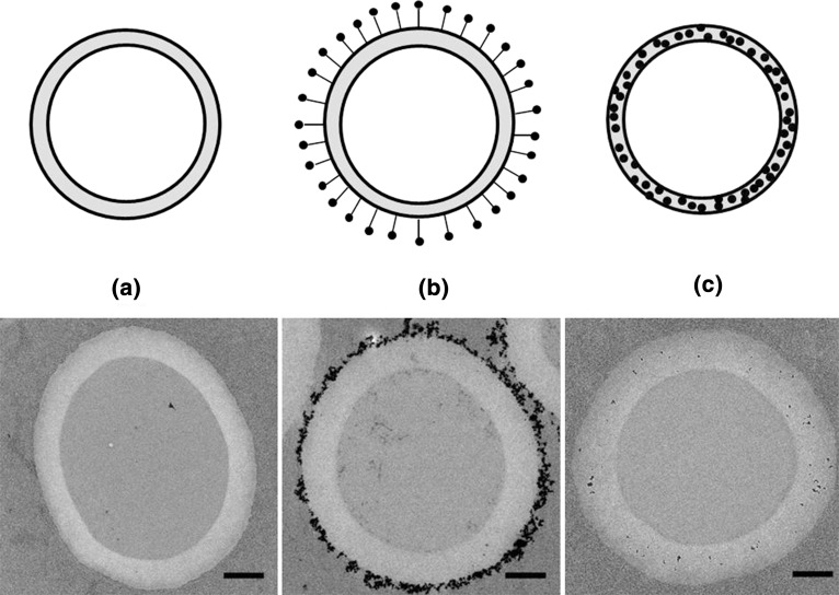
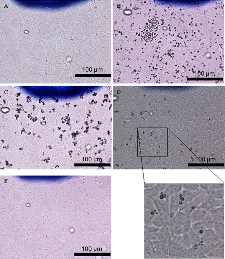
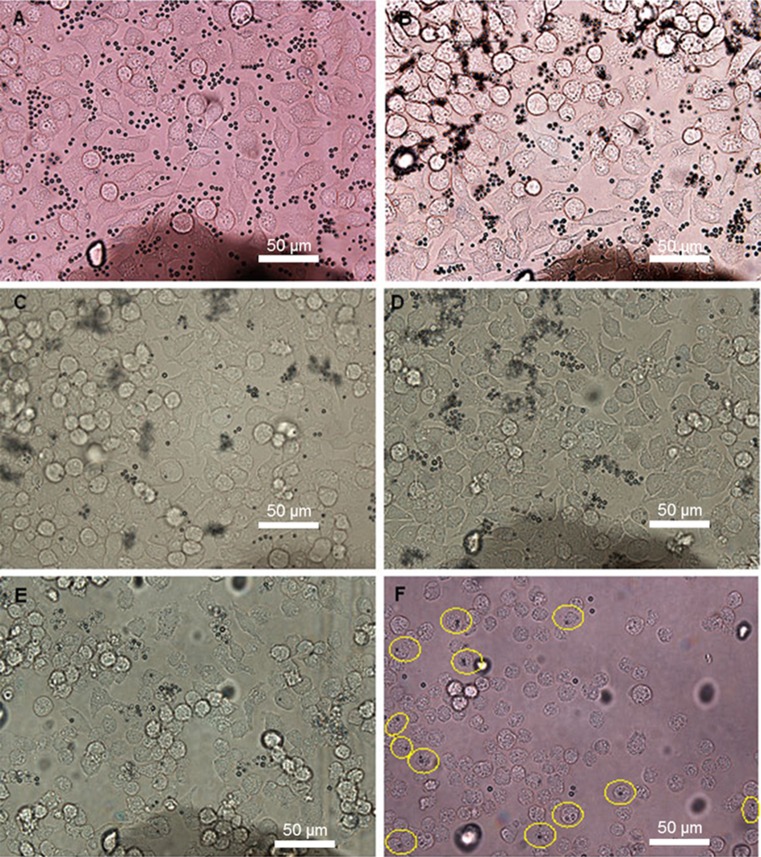
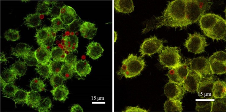
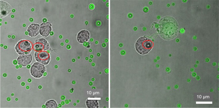
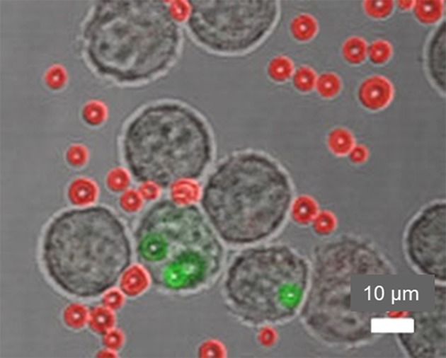
We studied the interactions between three types of non-targeted air-filled MBs with a polyvinyl-alcohol shell and murine macrophages or endothelial cells. The three MB types were plain MBs and two types that were labelled (internally and externally) with superparamagnetic iron oxide nanoparticles (SPIONs) for US/MRI bimodality. Cells were incubated with MBs and imaged by microscopy to evaluate uptake and adhesion. Interactions were quantified and the MB internalization was confirmed by fluorescence quenching of non-internalized MBs.
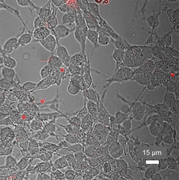
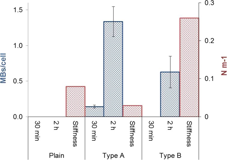
Macrophages internalized each MB type within different time frames: plain MBs 6 h, externally labelled MBs 25 min and internally labelled MBs 2 h. An average of 0.14 externally labelled MBs per cell were internalized after 30 min and 1.34 after 2 h; which was 113% more MBs than the number of internalized internally labelled MBs. The macrophages engulfed these three differently modified new MBs at various rate, whereas endothelial cells did not engulf MBs.
Polyvinyl-alcohol MBs are not taken up by endothelial cells. The MB uptake by macrophages is promoted by SPION labelling, in particular external such, which may be important for macrophage targeting.
通过使用对比增强技术可以提高诊断超声(US)和磁共振成像(MRI)的准确性。对于超声而言,充气微泡(MBs)或二氧化硅纳米颗粒(SiNPs),对于磁共振成像而言,超顺磁性或顺磁性剂都有助于实现这一点。然而,并非所有造影剂与血管壁和细胞的相互作用都已完全明确。
我们研究了三种带有聚乙烯醇外壳的非靶向充气微泡与小鼠巨噬细胞或内皮细胞之间的相互作用。这三种微泡类型分别是普通微泡以及两种用超顺磁性氧化铁纳米颗粒(SPIONs)进行(内部和外部)标记以实现超声/磁共振成像双模态的微泡。将细胞与微泡一起孵育,然后通过显微镜成像以评估摄取和黏附情况。对相互作用进行定量,并通过未内化微泡的荧光猝灭来确认微泡的内化。
巨噬细胞在不同时间内摄取每种微泡类型:普通微泡为6小时,外部标记的微泡为25分钟,内部标记的微泡为2小时。30分钟后每个细胞平均内化0.14个外部标记的微泡,2小时后为1.34个;这比内化的内部标记微泡数量多113%。巨噬细胞以不同速率吞噬这三种不同修饰的新型微泡,而内皮细胞不吞噬微泡。
聚乙烯醇微泡不被内皮细胞摄取。超顺磁性氧化铁纳米颗粒标记,特别是外部标记,可促进巨噬细胞对微泡的摄取,这对于巨噬细胞靶向可能很重要。