Bucci Domenico, Busceti Carla L, Calierno Maria T, Di Pietro Paola, Madonna Michele, Biagioni Francesca, Ryskalin Larisa, Limanaqi Fiona, Nicoletti Ferdinando, Fornai Francesco
Istituto Neurologico Mediterraneo (IRCCS), Neuromed, Pozzilli, Italy.
Department of Translational Research and New Technologies in Medicine and Surgery, University of Pisa, Pisa, Italy.
Front Neuroanat. 2017 Nov 2;11:98. doi: 10.3389/fnana.2017.00098. eCollection 2017.
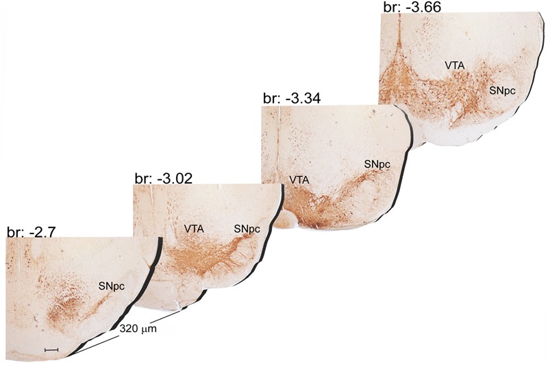
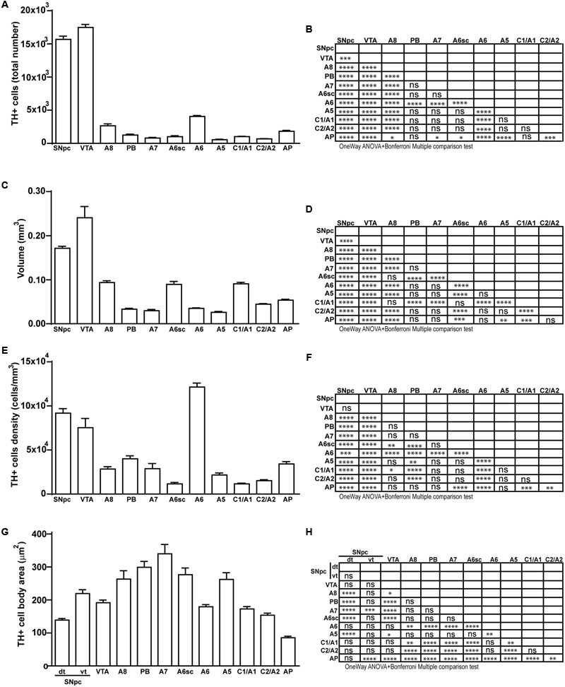
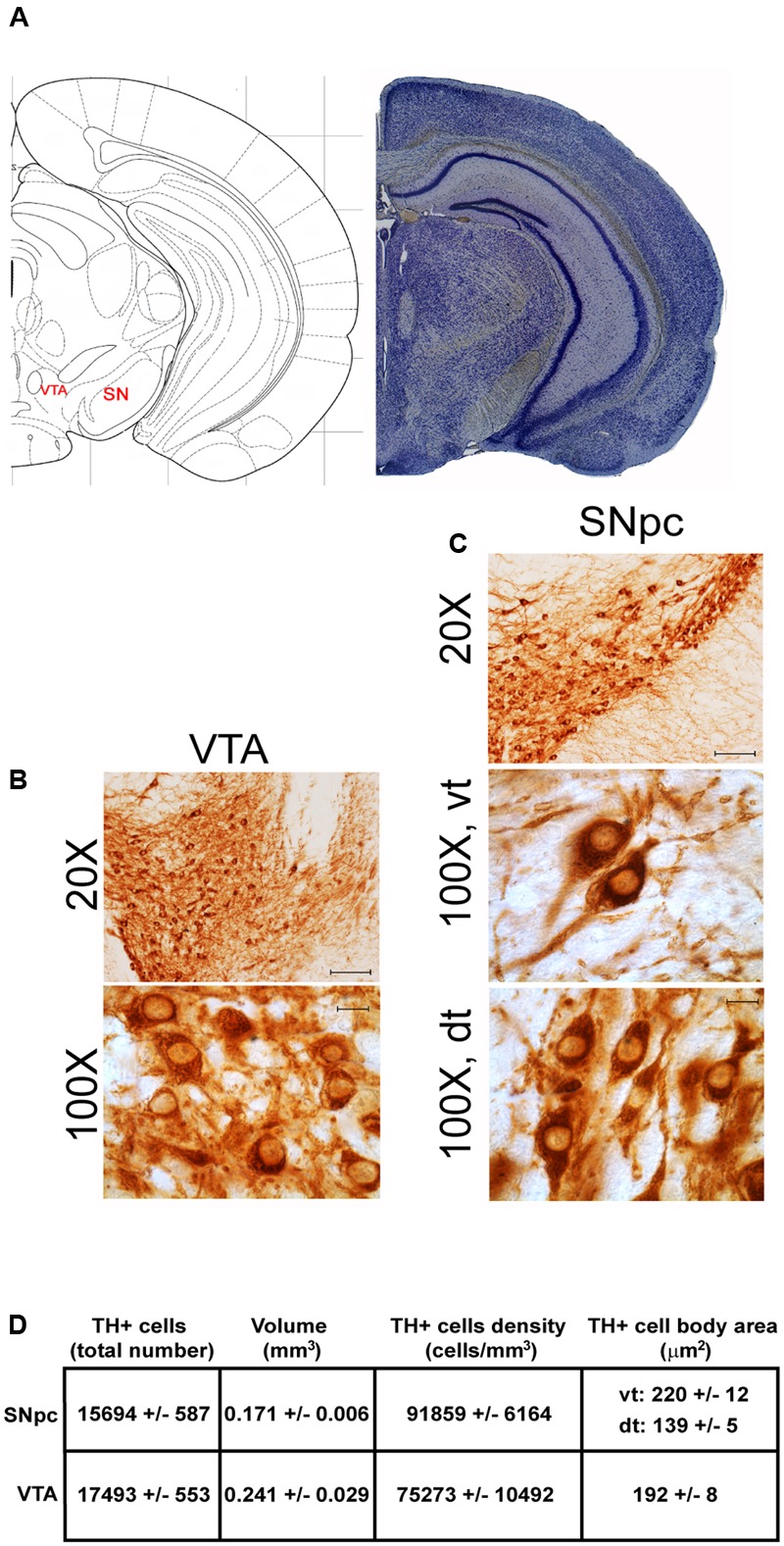
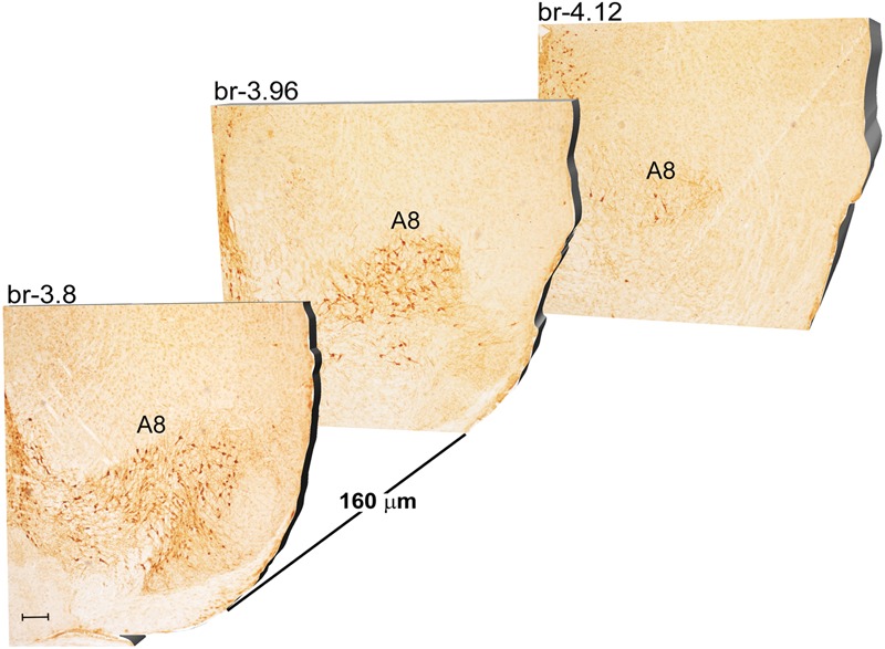
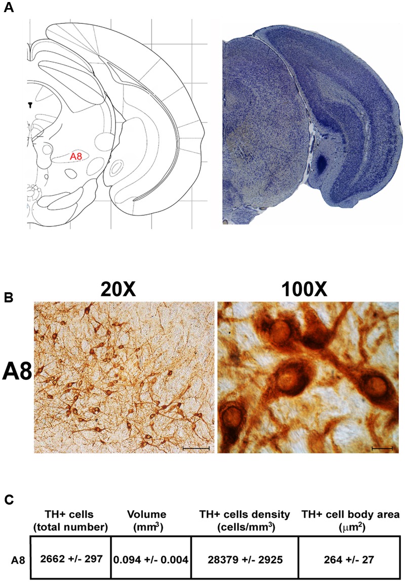
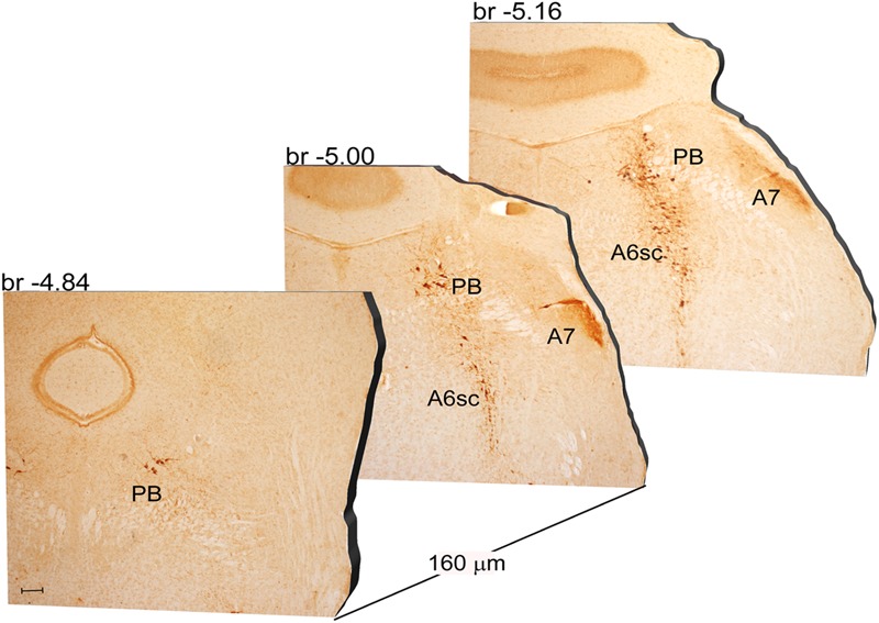
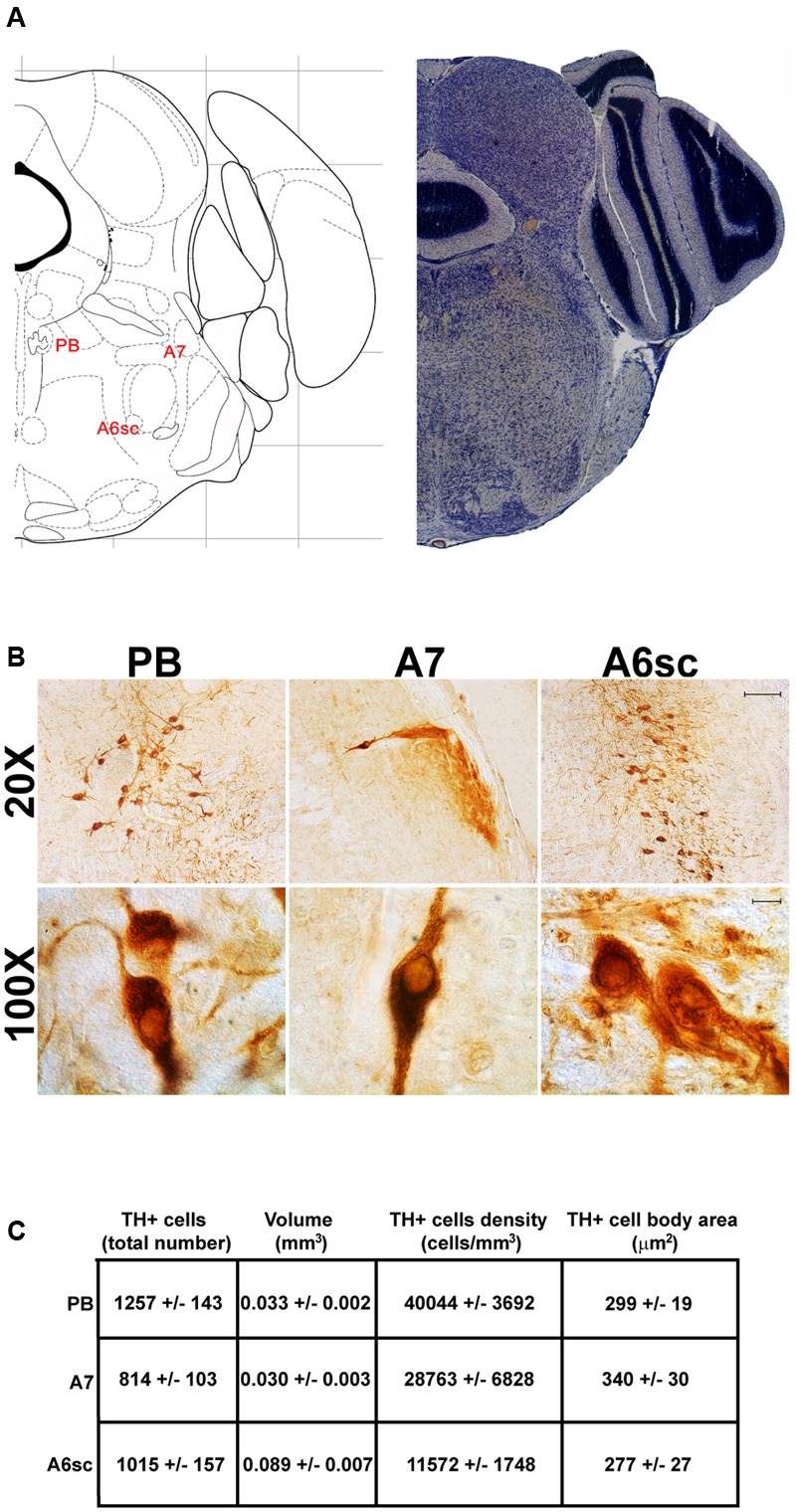
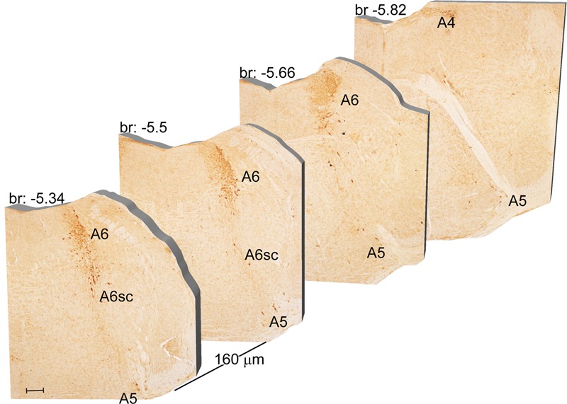
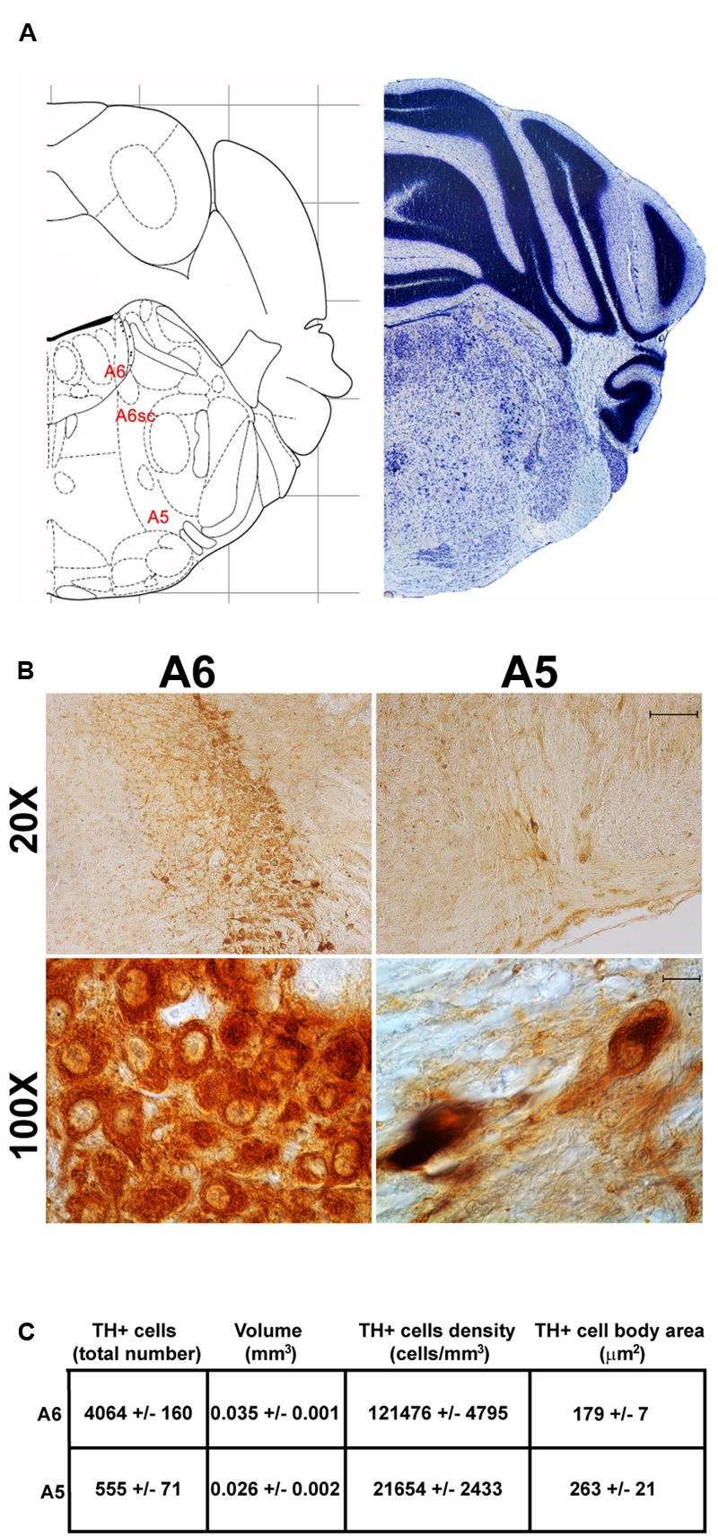
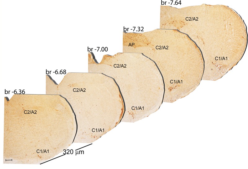
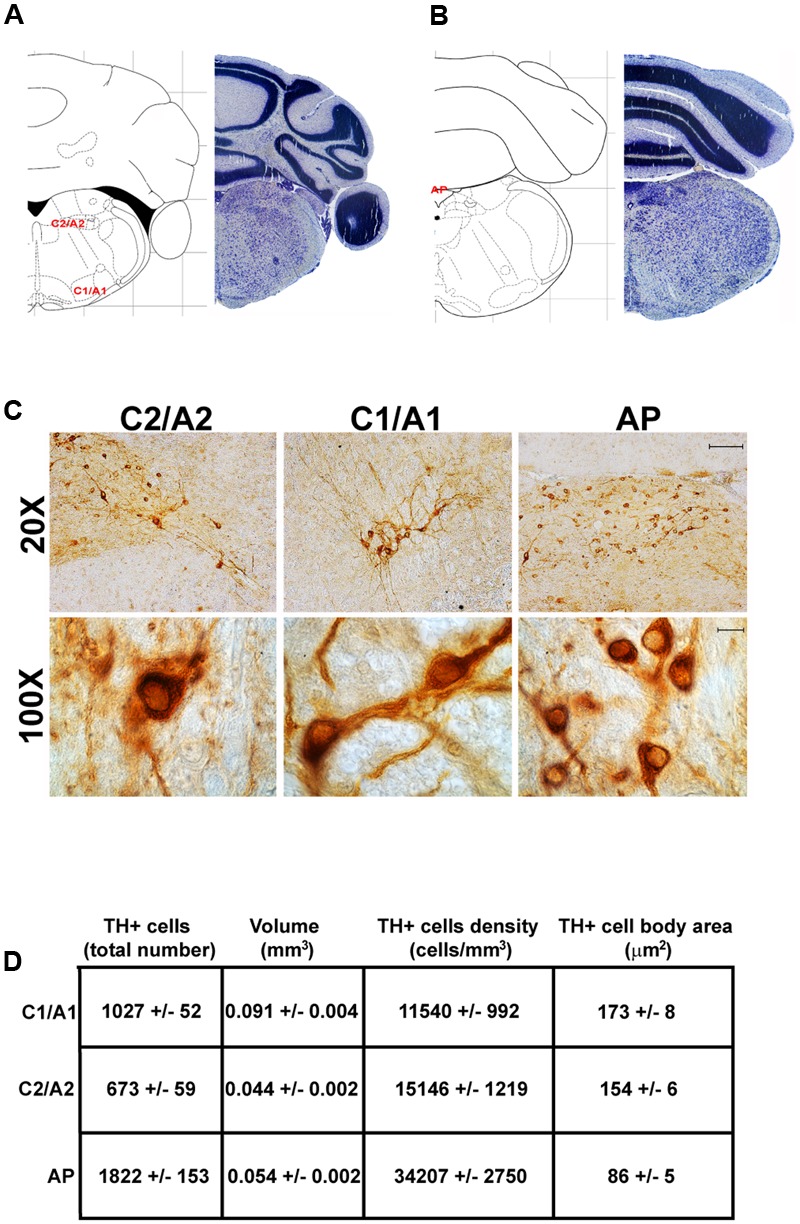
Catecholamine nuclei within the brainstem reticular formation (RF) play a pivotal role in a variety of brain functions. However, a systematic characterization of these nuclei in the very same experimental conditions is missing so far. Tyrosine hydroxylase (TH) immune-positive cells of the brainstem correspond to dopamine (DA)-, norepinephrine (NE)-, and epinephrine (E)-containing cells. Here, we report a systematic count of TH-positive neurons in the RF of the mouse brainstem by using stereological morphometry. All these nuclei were analyzed for anatomical localization, rostro-caudal extension, volume, neuron number, neuron density, and mean neuronal area for each nucleus. The present data apart from inherent informative value wish to represent a reference for neuronal mapping in those studies investigating the functional anatomy of the brainstem RF. These include: the sleep-wake cycle, movement control, muscle tone modulation, mood control, novelty orienting stimuli, attention, archaic responses to internal and external stressful stimuli, anxiety, breathing, blood pressure, and innumerable activities modulated by the archaic iso-dendritic hard core of the brainstem RF. Most TH-immune-positive cells fill the lateral part of the RF, which indeed possesses a high catecholamine content. A few nuclei are medial, although conventional nosography considers all these nuclei as part of the lateral column of the RF. Despite the key role of these nuclei in psychiatric and neurological disorders, only a few of them aspired a great attention in biomedical investigation, while most of them remain largely obscure although intense research is currently in progress. A simultaneous description of all these nuclei is not simply key to comprehend the variety of brainstem catecholamine reticular neurons, but probably represents an intrinsically key base for understanding brain physiology and physiopathology.
脑干网状结构(RF)中的儿茶酚胺核在多种脑功能中起关键作用。然而,迄今为止,在相同实验条件下对这些核进行系统表征的研究尚付阙如。脑干中酪氨酸羟化酶(TH)免疫阳性细胞对应于含多巴胺(DA)、去甲肾上腺素(NE)和肾上腺素(E)的细胞。在此,我们通过体视学形态测量法对小鼠脑干RF中TH阳性神经元进行了系统计数。对所有这些核的解剖定位、前后延伸、体积、神经元数量、神经元密度以及每个核的平均神经元面积进行了分析。本研究数据除了具有内在信息价值外,还希望为那些研究脑干RF功能解剖学的神经元图谱研究提供参考。这些功能包括:睡眠-觉醒周期、运动控制、肌张力调节、情绪控制、对新奇刺激的定向反应、注意力、对内外应激刺激的原始反应、焦虑、呼吸、血压,以及由脑干RF古老的同型树突硬核调节的无数活动。大多数TH免疫阳性细胞集中在RF的外侧部分,该部分确实含有高浓度的儿茶酚胺。有一些核位于内侧,尽管传统的疾病分类学将所有这些核视为RF外侧柱的一部分。尽管这些核在精神和神经疾病中起关键作用,但在生物医学研究中,只有少数核受到了极大关注,而大多数核尽管目前正在进行深入研究,但在很大程度上仍不清楚。同时描述所有这些核不仅是理解脑干儿茶酚胺网状神经元多样性的关键,而且可能是理解脑生理学和病理生理学的内在关键基础。