• 文献检索
  • 文档翻译
  • 深度研究
  • 学术资讯
  • Suppr Zotero 插件Zotero 插件
  • 邀请有礼
  • 套餐&价格
  • 历史记录
应用&插件
Suppr Zotero 插件Zotero 插件浏览器插件Mac 客户端Windows 客户端微信小程序
定价
高级版会员购买积分包购买API积分包
服务
文献检索文档翻译深度研究API 文档MCP 服务
关于我们
关于 Suppr公司介绍联系我们用户协议隐私条款
关注我们

Suppr 超能文献

核心技术专利:CN118964589B侵权必究
粤ICP备2023148730 号-1Suppr @ 2026

文献检索

告别复杂PubMed语法,用中文像聊天一样搜索,搜遍4000万医学文献。AI智能推荐,让科研检索更轻松。

立即免费搜索

文件翻译

保留排版,准确专业,支持PDF/Word/PPT等文件格式,支持 12+语言互译。

免费翻译文档

深度研究

AI帮你快速写综述,25分钟生成高质量综述,智能提取关键信息,辅助科研写作。

立即免费体验

视网膜脱离患者视觉通路区域自发性神经活动异常:一项静息态功能磁共振成像研究。

Abnormal regional spontaneous neural activity in visual pathway in retinal detachment patients: a resting-state functional MRI study.

作者信息

Huang Xin, Li Dan, Li Hai-Jun, Zhong Yu-Lin, Freeberg Shelby, Bao Jing, Zeng Xian-Jun, Shao Yi

机构信息

Department of Ophthalmology, The First Affiliated Hospital of Nanchang University, Jiangxi Province Clinical Ophthalmology Institute, Nanchang, Jiangxi, People's Republic of China.

Department of Ophthalmology, Eye Center, Renmin Hospital of Wuhan University, Wuhan University, Wuhan, Hubei, People's Republic of China.

出版信息

Neuropsychiatr Dis Treat. 2017 Nov 22;13:2849-2854. doi: 10.2147/NDT.S147645. eCollection 2017.

DOI:10.2147/NDT.S147645
PMID:29200859
原文链接:https://pmc.ncbi.nlm.nih.gov/articles/PMC5703148/
Abstract

OBJECTIVE

The aim of the study was to investigate changes of brain neural homogeneity in retinal detachment (RD) patients using the regional homogeneity (ReHo) method to understand their relationships with clinical features.

MATERIALS AND METHODS

A total of 30 patients with RD (16 men and 14 women), and 30 healthy controls (HCs) (16 men and 14 women) closely matched in age and sex were recruited. Resting-state functional magnetic resonance imaging scans were performed for all subjects. The ReHo method was used to investigate the brain regional neural homogeneity. Patients with RD were distinguished from HCs by receiver operating characteristic curve. The relationships between the mean ReHo signal values in many brain regions and clinical features in RD patients were calculated by Pearson correlation analysis.

RESULTS

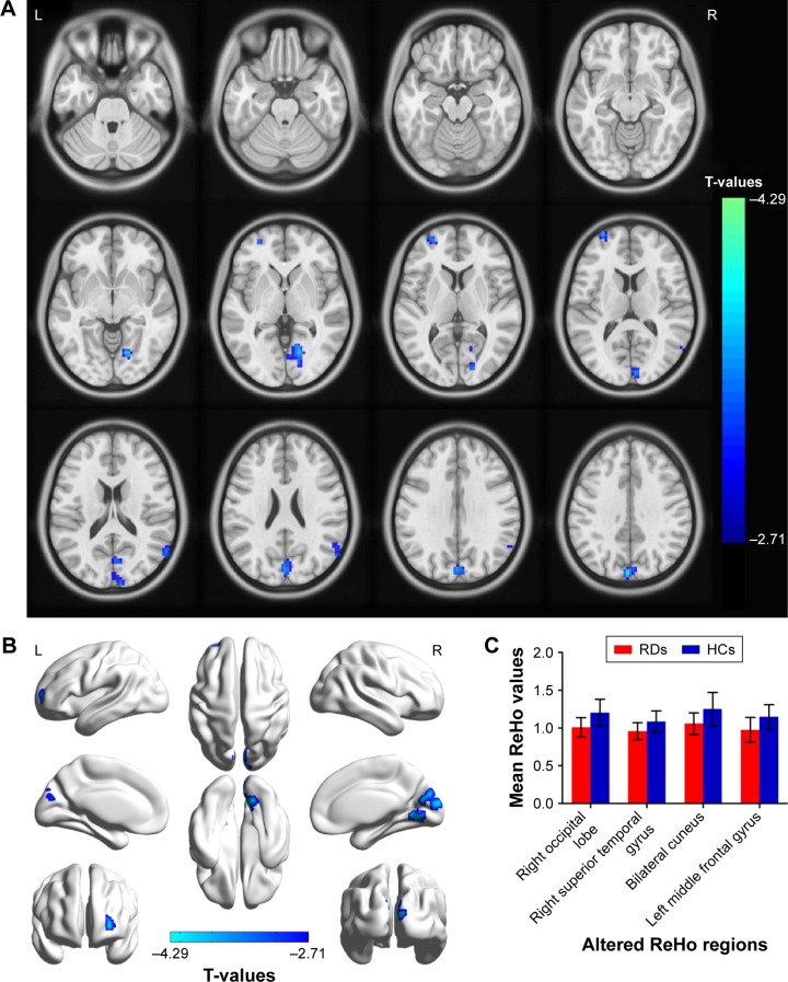
Compared with HCs, RD patients had significantly decreased ReHo values in the right occipital lobe, right superior temporal gyrus, bilateral cuneus and left middle frontal gyrus. Moreover, we found that the mean ReHo signal of the bilateral cuneus showed positive relationships with the duration of the RD (=0.392, =0.032).

CONCLUSION

The RD patients showed brain neural homogeneity dysfunction in the visual pathway, which may underline the pathological mechanism of RD patients with acute vision loss. Besides, the ReHo values can reflect the progress of the RD disease.

摘要

目的

本研究旨在采用局部一致性(ReHo)方法研究视网膜脱离(RD)患者脑神经网络同质性的变化,以了解其与临床特征的关系。

材料与方法

招募30例RD患者(男16例,女14例)和30例健康对照者(HCs)(男16例,女14例),年龄和性别相匹配。对所有受试者进行静息态功能磁共振成像扫描。采用ReHo方法研究脑区神经同质性。通过受试者工作特征曲线将RD患者与HCs区分开来。采用Pearson相关分析计算RD患者多个脑区平均ReHo信号值与临床特征之间的关系。

结果

与HCs相比,RD患者右侧枕叶、右侧颞上回、双侧楔叶和左侧额中回的ReHo值显著降低。此外,我们发现双侧楔叶的平均ReHo信号与RD持续时间呈正相关(r = 0.392,P = 0.032)。

结论

RD患者在视觉通路中存在脑神经网络同质性功能障碍,这可能是RD患者急性视力丧失的病理机制。此外,ReHo值可以反映RD疾病的进展。

https://cdn.ncbi.nlm.nih.gov/pmc/blobs/95f8/5703148/f272f717d5f7/ndt-13-2849Fig3.jpg
https://cdn.ncbi.nlm.nih.gov/pmc/blobs/95f8/5703148/38b0a2fc9dd8/ndt-13-2849Fig1.jpg
https://cdn.ncbi.nlm.nih.gov/pmc/blobs/95f8/5703148/c240e86076a9/ndt-13-2849Fig2.jpg
https://cdn.ncbi.nlm.nih.gov/pmc/blobs/95f8/5703148/f272f717d5f7/ndt-13-2849Fig3.jpg
https://cdn.ncbi.nlm.nih.gov/pmc/blobs/95f8/5703148/38b0a2fc9dd8/ndt-13-2849Fig1.jpg
https://cdn.ncbi.nlm.nih.gov/pmc/blobs/95f8/5703148/c240e86076a9/ndt-13-2849Fig2.jpg
https://cdn.ncbi.nlm.nih.gov/pmc/blobs/95f8/5703148/f272f717d5f7/ndt-13-2849Fig3.jpg

相似文献

1
Abnormal regional spontaneous neural activity in visual pathway in retinal detachment patients: a resting-state functional MRI study.视网膜脱离患者视觉通路区域自发性神经活动异常:一项静息态功能磁共振成像研究。
Neuropsychiatr Dis Treat. 2017 Nov 22;13:2849-2854. doi: 10.2147/NDT.S147645. eCollection 2017.
2
Comparison of spontaneous brain activity revealed by regional homogeneity in AQP4-IgG neuromyelitis optica-optic neuritis versus MOG-IgG optic neuritis patients: a resting-state functional MRI study.水通道蛋白4-IgG视神经脊髓炎-视神经炎与髓鞘少突胶质细胞糖蛋白-IgG视神经炎患者中基于局部一致性揭示的自发脑活动比较:一项静息态功能磁共振成像研究
Neuropsychiatr Dis Treat. 2017 Oct 24;13:2669-2679. doi: 10.2147/NDT.S145183. eCollection 2017.
3
ALTERED BRAIN ACTIVITY IN PATIENTS WITH DIABETIC RETINOPATHY USING REGIONAL HOMOGENEITY: A RESTING-STATE fMRI STUDY.糖尿病视网膜病变患者局部一致性的脑活动改变:一项静息态 fMRI 研究。
Endocr Pract. 2019 Apr;25(4):320-327. doi: 10.4158/EP-2018-0517.
4
Functional connectivity density alterations in middle-age retinal detachment patients.中年视网膜脱离患者的功能连接密度改变。
Brain Behav. 2021 Apr;11(4):e01783. doi: 10.1002/brb3.1783. Epub 2021 Mar 1.
5
[Relationship between auditory hallucination and regional homogeneity of functional magnetic resonance imaging in first-episode childhood and adolescence-onset schizophrenia].首发儿童及青少年期精神分裂症幻听与功能磁共振成像局部一致性的关系
Zhonghua Yi Xue Za Zhi. 2021 Jun 29;101(24):1915-1920. doi: 10.3760/cma.j.cn112137-20201126-03195.
6
Altered regional homogeneity in patients with late monocular blindness: a resting-state functional MRI study.晚期单眼失明患者的局部一致性改变:一项静息态功能磁共振成像研究
Neuroreport. 2017 Nov 8;28(16):1085-1091. doi: 10.1097/WNR.0000000000000855.
7
Altered Regional Homogeneity in Patients With Corneal Ulcer: A Resting-State Functional MRI Study.角膜溃疡患者的局部一致性改变:一项静息态功能磁共振成像研究
Front Neurosci. 2019 Jul 23;13:743. doi: 10.3389/fnins.2019.00743. eCollection 2019.
8
Altered regional homogeneity in patients with unilateral acute open-globe injury: a resting-state functional MRI study.单侧急性开放性眼球损伤患者的局部一致性改变:一项静息态功能磁共振成像研究。
Neuropsychiatr Dis Treat. 2016 Aug 1;12:1901-6. doi: 10.2147/NDT.S110541. eCollection 2016.
9
[Diffusion tensor imaging and resting-state functional magnetic resonance imaging in patients with delirium in intensive care unit].[重症监护病房谵妄患者的扩散张量成像与静息态功能磁共振成像]
Zhonghua Wei Zhong Bing Ji Jiu Yi Xue. 2020 Jan;32(1):88-93. doi: 10.3760/cma.j.cn121430-20190905-00016.
10
[Regional homogeneity in the patients of irritable bowel syndrome complicated with depression: a resting-state functional magnetic resonance imaging study].肠易激综合征合并抑郁症患者的局部一致性:一项静息态功能磁共振成像研究
Zhonghua Yi Xue Za Zhi. 2018 Jan 16;98(3):196-201. doi: 10.3760/cma.j.issn.0376-2491.2018.03.008.

引用本文的文献

1
Guidelines for preoperative visual function and imaging examination standards in vitreoretinal surgery (2025).玻璃体视网膜手术术前视觉功能及影像学检查标准指南(2025年)
Int J Ophthalmol. 2025 May 18;18(5):813-831. doi: 10.18240/ijo.2025.05.06. eCollection 2025.
2
Abnormal intrinsic brain functional network dynamics in patients with retinal detachment based on graph theory and machine learning.基于图论和机器学习的视网膜脱离患者大脑内在功能网络动力学异常
Heliyon. 2024 Nov 2;10(23):e37890. doi: 10.1016/j.heliyon.2024.e37890. eCollection 2024 Dec 15.
3
OCTA evaluates changes in retinal microvasculature in renal hypertension patients.

本文引用的文献

1
Retinal Detachment Associated With Basketball-Related Eye Trauma.与篮球相关的眼部创伤所致视网膜脱离
Am J Ophthalmol. 2017 Aug;180:97-101. doi: 10.1016/j.ajo.2017.05.025. Epub 2017 Jun 21.
2
Retinal detachment surgery in Western Australia (2000-2013): a whole-population study.西澳大利亚州的视网膜脱离手术(2000 - 2013年):一项全人群研究。
Br J Ophthalmol. 2017 Dec;101(12):1679-1682. doi: 10.1136/bjophthalmol-2016-310070. Epub 2017 Apr 8.
3
Altered intrinsic regional brain spontaneous activity in patients with comitant strabismus: a resting-state functional MRI study.
OCTA 评估肾性高血压患者视网膜微血管的变化。
Sci Rep. 2024 Nov 21;14(1):28910. doi: 10.1038/s41598-024-68690-3.
4
Machine learning analysis reveals aberrant dynamic changes in amplitude of low-frequency fluctuations among patients with retinal detachment.机器学习分析揭示了视网膜脱离患者低频波动幅度的异常动态变化。
Front Neurosci. 2023 Jul 20;17:1227081. doi: 10.3389/fnins.2023.1227081. eCollection 2023.
5
Abnormal percent amplitude of fluctuation changes in patients with monocular blindness: A resting-state functional magnetic resonance imaging study.单眼失明患者波动变化的异常百分比幅度:一项静息态功能磁共振成像研究。
Front Psychiatry. 2022 Oct 24;13:942905. doi: 10.3389/fpsyt.2022.942905. eCollection 2022.
6
Reduced cortical complexity in patients with thyroid-associated ophthalmopathy.甲状腺相关性眼病患者皮质复杂性降低。
Brain Imaging Behav. 2022 Oct;16(5):2133-2140. doi: 10.1007/s11682-022-00683-0. Epub 2022 Jul 12.
7
Altered Regional Homogeneity in Patients With Congenital Blindness: A Resting-State Functional Magnetic Resonance Imaging Study.先天性失明患者的局部一致性改变:一项静息态功能磁共振成像研究。
Front Psychiatry. 2022 Jun 22;13:925412. doi: 10.3389/fpsyt.2022.925412. eCollection 2022.
8
Altered Brain Activity in Strabismic Amblyopic Children as Determined by Regional Homogeneity: A Resting-State Functional Magnetic Resonance Imaging Study.基于局部一致性测定的斜视性弱视儿童脑活动改变:一项静息态功能磁共振成像研究
Front Neurosci. 2022 Jun 2;16:879253. doi: 10.3389/fnins.2022.879253. eCollection 2022.
9
Brain Activity in Age-Related Macular Degeneration Patients From the Perspective of Regional Homogeneity: A Resting-State Functional Magnetic Resonance Imaging Study.基于局部一致性视角的年龄相关性黄斑变性患者脑活动:一项静息态功能磁共振成像研究
Front Aging Neurosci. 2022 May 9;14:865430. doi: 10.3389/fnagi.2022.865430. eCollection 2022.
10
Local-to-Remote Brain Functional Connectivity in Patients with Thyroid-Associated Ophthalmopathy and Assessment of Its Predictive Value Using Machine Learning.甲状腺相关性眼病患者的局部到远程脑功能连接及其机器学习预测价值评估
Int J Gen Med. 2022 Apr 21;15:4273-4283. doi: 10.2147/IJGM.S353649. eCollection 2022.
共同性斜视患者脑内固有区域自发活动的改变:一项静息态功能磁共振成像研究
Neuropsychiatr Dis Treat. 2016 Jun 3;12:1303-8. doi: 10.2147/NDT.S105478. eCollection 2016.
4
Altered intrinsic regional spontaneous brain activity in patients with optic neuritis: a resting-state functional magnetic resonance imaging study.视神经炎患者脑内固有区域自发脑活动的改变:一项静息态功能磁共振成像研究
Neuropsychiatr Dis Treat. 2015 Dec 11;11:3065-73. doi: 10.2147/NDT.S92968. eCollection 2015.
5
Loss of Visual Acuity after Successful Surgery for Macula-On Rhegmatogenous Retinal Detachment in a Prospective Multicentre Study.一项前瞻性多中心研究:孔源性黄斑脱离成功手术后的视力丧失
J Ophthalmol. 2015;2015:821864. doi: 10.1155/2015/821864. Epub 2015 Nov 12.
6
Anatomical pathways for auditory memory II: information from rostral superior temporal gyrus to dorsolateral temporal pole and medial temporal cortex.听觉记忆的解剖学通路II:从颞上回前部到颞极背外侧和颞叶内侧皮质的信息
Front Neurosci. 2015 May 18;9:158. doi: 10.3389/fnins.2015.00158. eCollection 2015.
7
Ageing of the vitreous: From acute onset floaters and flashes to retinal detachment.玻璃体老化:从急性飞蚊症和闪光到视网膜脱离。
Ageing Res Rev. 2015 May;21:71-7. doi: 10.1016/j.arr.2015.03.006. Epub 2015 Apr 2.
8
Cuneus and fusiform cortices thickness is reduced in trigeminal neuralgia.三叉神经痛患者楔叶和梭状回皮质厚度减少。
J Headache Pain. 2014 Mar 24;15(1):17. doi: 10.1186/1129-2377-15-17.
9
Bilateral retinal detachment with subsequent blindness in a pregnant woman with severe pre-eclampsia.一名患有重度子痫前期的孕妇发生双侧视网膜脱离并随后失明。
Taiwan J Obstet Gynecol. 2013 Mar;52(1):142-4. doi: 10.1016/j.tjog.2012.05.001.
10
Decreased regional homogeneity in lingual gyrus, increased regional homogeneity in cuneus and correlations with panic symptom severity of first-episode, medication-naïve and late-onset panic disorder patients.首发、未经药物治疗和晚发性惊恐障碍患者舌回局部一致性降低,楔叶局部一致性增加,且与惊恐症状严重程度呈正相关。
Psychiatry Res. 2013 Feb 28;211(2):127-31. doi: 10.1016/j.pscychresns.2012.11.006. Epub 2013 Jan 24.