• 文献检索
  • 文档翻译
  • 深度研究
  • 学术资讯
  • Suppr Zotero 插件Zotero 插件
  • 邀请有礼
  • 套餐&价格
  • 历史记录
应用&插件
Suppr Zotero 插件Zotero 插件浏览器插件Mac 客户端Windows 客户端微信小程序
定价
高级版会员购买积分包购买API积分包
服务
文献检索文档翻译深度研究API 文档MCP 服务
关于我们
关于 Suppr公司介绍联系我们用户协议隐私条款
关注我们

Suppr 超能文献

核心技术专利:CN118964589B侵权必究
粤ICP备2023148730 号-1Suppr @ 2026

文献检索

告别复杂PubMed语法,用中文像聊天一样搜索,搜遍4000万医学文献。AI智能推荐,让科研检索更轻松。

立即免费搜索

文件翻译

保留排版,准确专业,支持PDF/Word/PPT等文件格式,支持 12+语言互译。

免费翻译文档

深度研究

AI帮你快速写综述,25分钟生成高质量综述,智能提取关键信息,辅助科研写作。

立即免费体验

成年大鼠脊髓局灶性损伤后经典活化小胶质细胞移植对白质再生影响的磁共振成像扩展研究

Extended magnetic resonance imaging studies on the effect of classically activated microglia transplantation on white matter regeneration following spinal cord focal injury in adult rats.

作者信息

Marcol Wiesław, Ślusarczyk Wojciech, Larysz-Brysz Magdalena, Łabuzek Krzysztof, Kapustka Bartosz, Staszkiewicz Rafał, Rosicka Paulina, Kalita Katarzyna, Węglarz Władysław, Lewin-Kowalik Joanna

机构信息

Department of Physiology, School of Medicine in Katowice, Medical University of Silesia, 40-752 Katowice, Poland.

Department of Internal Medicine and Clinical Pharmacology, School of Medicine in Katowice, Medical University of Silesia, 40-752 Katowice, Poland.

出版信息

Exp Ther Med. 2017 Nov;14(5):4869-4877. doi: 10.3892/etm.2017.5130. Epub 2017 Sep 19.

DOI:10.3892/etm.2017.5130
PMID:29201191
原文链接:https://pmc.ncbi.nlm.nih.gov/articles/PMC5704303/
Abstract

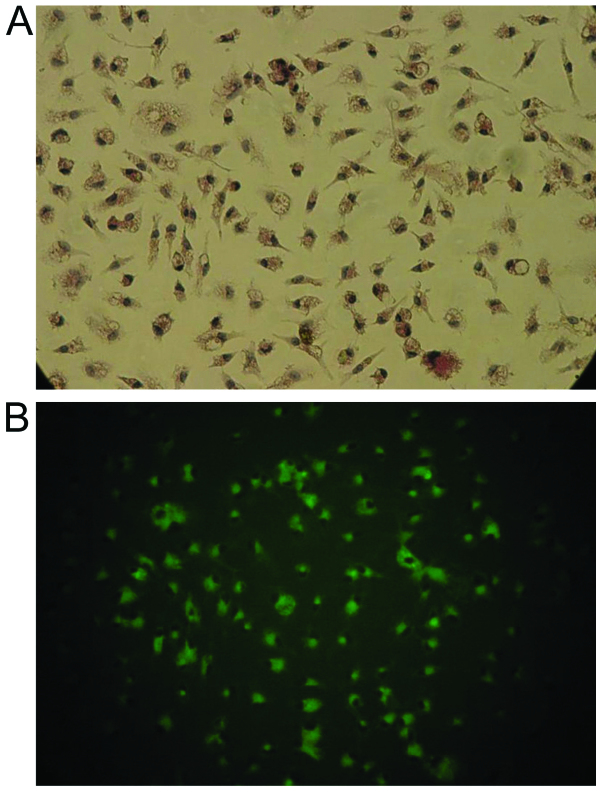
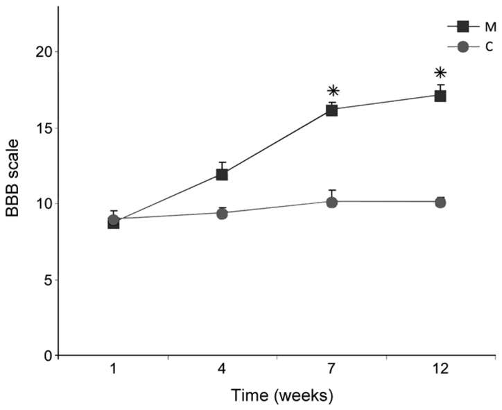
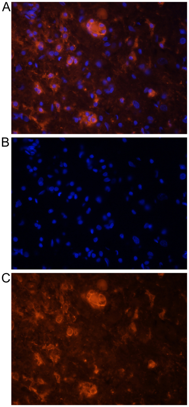
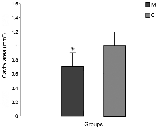
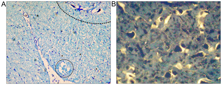
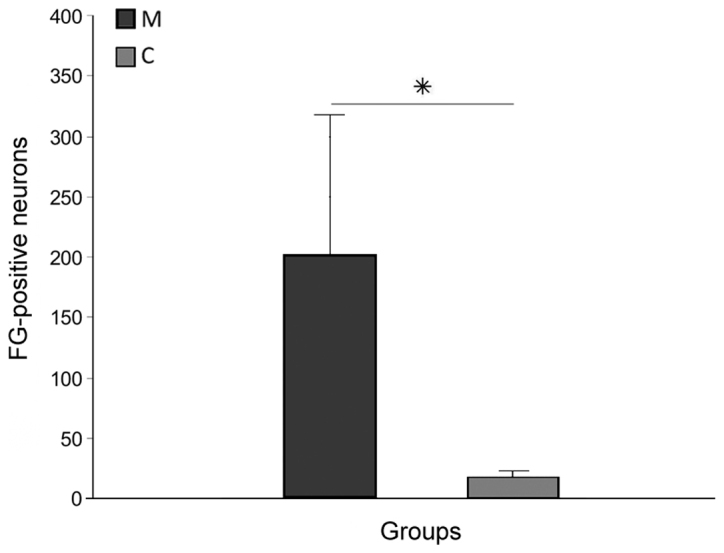
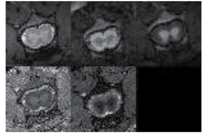
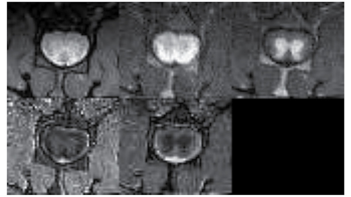
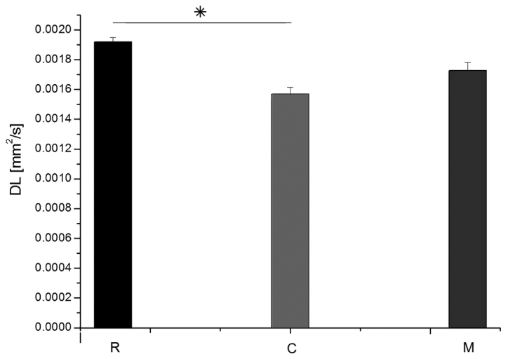
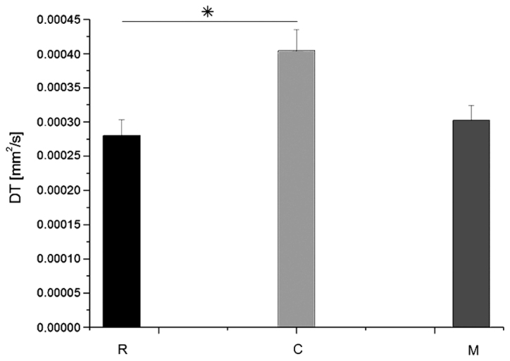
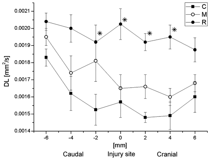
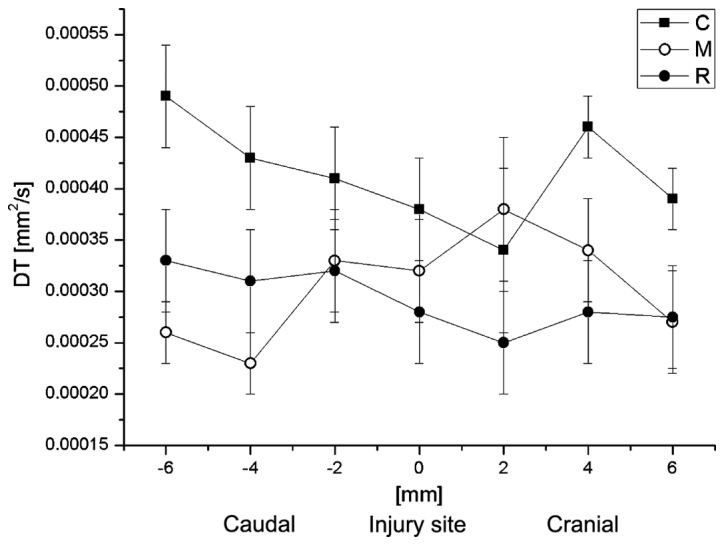
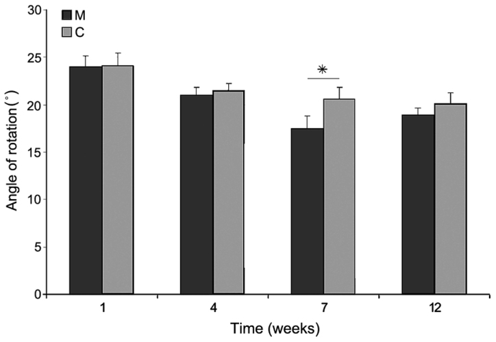
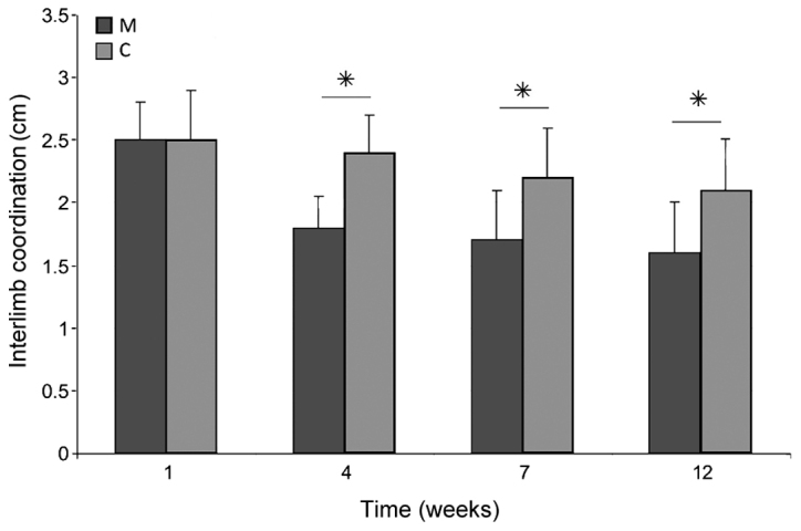
Spinal cord injuries are still a serious problem for regenerative medicine. Previous research has demonstrated that activated microglia accumulate in spinal lesions, influencing the injured tissues in various ways. Therefore, transplantation of activated microglia may have a beneficial role in the regeneration of the nervous system. The present study examined the influence of transplanted activated microglial cells in adult rats with injured spinal cords. Rats were randomly divided into an experimental (M) and control (C) group, and were subjected to non-laminectomy focal injury of spinal cord white matter by means of a high-pressured air stream. In group M, activated cultured microglial cells were injected twice into the site of injury. Functional outcome and morphological features of regeneration were analyzed during a 12-week follow-up. The lesions were characterized by means of magnetic resonance imaging (MRI). Neurons in the brain stem and motor cortex were labeled with FluoroGold (FG). A total of 12 weeks after surgery, spinal cords and brains were collected and subjected to histopathological and immunohistochemical examinations. Lesion sizes in the spinal cord were measured and the number of FG-positive neurons was counted. Rats in group M demonstrated significant improvement of locomotor performance when compared with group C (P<0.05). MRI analysis demonstrated moderate improvement in water diffusion along the spinal cord in the group M following microglia treatment, as compared with group C. The water diffusion perpendicular to the spinal cord in group M was closer to the reference values for a healthy spinal cord than it was in group C. The sizes of lesions were also significantly smaller in group M than in the group C (P<0.05). The number of brain stem and motor cortex FG-positive neurons in group M was significantly higher than in group C. The present study demonstrated that delivery of activated microglia directly into the injured spinal cord gives some positive effects for the regeneration of the white matter.

摘要

脊髓损伤对于再生医学来说仍然是一个严重的问题。先前的研究表明,活化的小胶质细胞在脊髓损伤部位积聚,以多种方式影响受损组织。因此,移植活化的小胶质细胞可能对神经系统的再生具有有益作用。本研究考察了移植活化的小胶质细胞对成年脊髓损伤大鼠的影响。大鼠被随机分为实验组(M组)和对照组(C组),通过高压气流对脊髓白质进行非椎板切除术局灶性损伤。在M组中,将培养的活化小胶质细胞分两次注射到损伤部位。在12周的随访期间分析再生的功能结果和形态特征。通过磁共振成像(MRI)对损伤进行表征。用荧光金(FG)标记脑干和运动皮层中的神经元。手术后共12周,收集脊髓和大脑并进行组织病理学和免疫组织化学检查。测量脊髓损伤大小并计数FG阳性神经元的数量。与C组相比,M组大鼠的运动性能有显著改善(P<0.05)。MRI分析表明,与C组相比,M组在小胶质细胞治疗后脊髓水扩散有中度改善。M组垂直于脊髓的水扩散比C组更接近健康脊髓的参考值。M组的损伤大小也明显小于C组(P<0.05)。M组脑干和运动皮层FG阳性神经元的数量明显高于C组。本研究表明,将活化的小胶质细胞直接输送到受损脊髓中对白质再生有一些积极作用。

https://cdn.ncbi.nlm.nih.gov/pmc/blobs/2c2b/5704303/1f3a03f403b1/etm-14-05-4869-g13.jpg
https://cdn.ncbi.nlm.nih.gov/pmc/blobs/2c2b/5704303/b735ac2720ae/etm-14-05-4869-g00.jpg
https://cdn.ncbi.nlm.nih.gov/pmc/blobs/2c2b/5704303/e62279fa8948/etm-14-05-4869-g01.jpg
https://cdn.ncbi.nlm.nih.gov/pmc/blobs/2c2b/5704303/ed0f453215bb/etm-14-05-4869-g02.jpg
https://cdn.ncbi.nlm.nih.gov/pmc/blobs/2c2b/5704303/ff31bb9f710c/etm-14-05-4869-g03.jpg
https://cdn.ncbi.nlm.nih.gov/pmc/blobs/2c2b/5704303/daeac2be827e/etm-14-05-4869-g04.jpg
https://cdn.ncbi.nlm.nih.gov/pmc/blobs/2c2b/5704303/1ceea2e884e2/etm-14-05-4869-g05.jpg
https://cdn.ncbi.nlm.nih.gov/pmc/blobs/2c2b/5704303/d3b5a180b650/etm-14-05-4869-g06.jpg
https://cdn.ncbi.nlm.nih.gov/pmc/blobs/2c2b/5704303/28c874df2a36/etm-14-05-4869-g07.jpg
https://cdn.ncbi.nlm.nih.gov/pmc/blobs/2c2b/5704303/f4da7c017e98/etm-14-05-4869-g08.jpg
https://cdn.ncbi.nlm.nih.gov/pmc/blobs/2c2b/5704303/13472569e751/etm-14-05-4869-g09.jpg
https://cdn.ncbi.nlm.nih.gov/pmc/blobs/2c2b/5704303/0640abd8991a/etm-14-05-4869-g10.jpg
https://cdn.ncbi.nlm.nih.gov/pmc/blobs/2c2b/5704303/abbbbff2e02e/etm-14-05-4869-g11.jpg
https://cdn.ncbi.nlm.nih.gov/pmc/blobs/2c2b/5704303/fc370ef8b8d6/etm-14-05-4869-g12.jpg
https://cdn.ncbi.nlm.nih.gov/pmc/blobs/2c2b/5704303/1f3a03f403b1/etm-14-05-4869-g13.jpg

相似文献

1
Extended magnetic resonance imaging studies on the effect of classically activated microglia transplantation on white matter regeneration following spinal cord focal injury in adult rats.成年大鼠脊髓局灶性损伤后经典活化小胶质细胞移植对白质再生影响的磁共振成像扩展研究
Exp Ther Med. 2017 Nov;14(5):4869-4877. doi: 10.3892/etm.2017.5130. Epub 2017 Sep 19.
2
Bone marrow stem cells delivered into the subarachnoid space via cisterna magna improve repair of injured rat spinal cord white matter.经枕大池将骨髓干细胞注入蛛网膜下腔可改善大鼠脊髓损伤白质的修复。
Int J Clin Exp Med. 2015 Sep 15;8(9):14680-92. eCollection 2015.
3
Grafted Activated Schwann Cells Support Survival of Injured Rat Spinal Cord White Matter.移植的活化雪旺细胞支持损伤大鼠脊髓白质的存活。
World Neurosurg. 2015 Aug;84(2):511-9. doi: 10.1016/j.wneu.2015.04.027. Epub 2015 Apr 21.
4
The neuroprotective effect of N-acetylcysteine in spinal cord-injured rats.N-乙酰半胱氨酸对脊髓损伤大鼠的神经保护作用。
Adv Clin Exp Med. 2017 Dec;26(9):1329-1334. doi: 10.17219/acem/65478.
5
Functional recovery after human umbilical cord blood cells transplantation with brain-derived neutrophic factor into the spinal cord injured rat.将人脐带血细胞与脑源性神经营养因子一同移植到脊髓损伤大鼠体内后的功能恢复情况
Acta Neurochir (Wien). 2005 Sep;147(9):985-92; discussion 992. doi: 10.1007/s00701-005-0538-y. Epub 2005 Jul 11.
6
Differential activation of astrocytes and microglia after spinal cord injury in the fetal rat.胎鼠脊髓损伤后星形胶质细胞和小胶质细胞的差异性激活
Eur Spine J. 2006 Feb;15(2):223-33. doi: 10.1007/s00586-005-0933-3. Epub 2005 Nov 16.
7
Human neural stem cells promote corticospinal axons regeneration and synapse reformation in injured spinal cord of rats.人神经干细胞促进大鼠脊髓损伤后皮质脊髓轴突再生和突触重塑。
Chin Med J (Engl). 2006 Aug 20;119(16):1331-8.
8
Transplantation of oligodendrocyte precursors and sonic hedgehog results in improved function and white matter sparing in the spinal cords of adult rats after contusion.少突胶质前体细胞移植与音猬因子共同作用可改善成年大鼠脊髓挫伤后的功能并减少白质损伤。
Spine J. 2004 Jan-Feb;4(1):16-26. doi: 10.1016/j.spinee.2003.07.004.
9
Effects of early surgical decompression on functional and histological outcomes after severe experimental thoracic spinal cord injury.早期手术减压对严重实验性胸段脊髓损伤后功能和组织学结果的影响。
J Neurosurg Spine. 2017 Jan;26(1):62-75. doi: 10.3171/2016.6.SPINE16343. Epub 2016 Sep 16.
10
Cell cycle inhibition attenuates microglia induced inflammatory response and alleviates neuronal cell death after spinal cord injury in rats.细胞周期抑制可减轻大鼠脊髓损伤后小胶质细胞诱导的炎症反应并减轻神经元细胞死亡。
Brain Res. 2007 Mar 2;1135(1):177-85. doi: 10.1016/j.brainres.2006.11.085. Epub 2006 Dec 26.

引用本文的文献

1
Transplantation of M2-Deviated Microglia Promotes Recovery of Motor Function after Spinal Cord Injury in Mice.M2 型小胶质细胞移植促进小鼠脊髓损伤后运动功能的恢复。
Mol Ther. 2020 Jan 8;28(1):254-265. doi: 10.1016/j.ymthe.2019.09.004. Epub 2019 Sep 10.
2
Use of ebselen as a neuroprotective agent in rat spinal cord subjected to traumatic injury.依布硒啉作为神经保护剂在遭受创伤性损伤的大鼠脊髓中的应用。
Neural Regen Res. 2019 Jul;14(7):1255-1261. doi: 10.4103/1673-5374.251334.

本文引用的文献

1
Improved regeneration after spinal cord injury in mice lacking functional T- and B-lymphocytes.T 细胞和 B 细胞功能缺失的小鼠脊髓损伤后再生能力增强。
Exp Neurol. 2012 Oct;237(2):274-85. doi: 10.1016/j.expneurol.2012.07.016. Epub 2012 Jul 31.
2
Bone marrow-derived stromal cell therapy for traumatic brain injury is neuroprotective via stimulation of non-neurologic organ systems.骨髓基质细胞治疗创伤性脑损伤是通过刺激非神经系统器官系统起神经保护作用的。
Surgery. 2012 Nov;152(5):790-3. doi: 10.1016/j.surg.2012.06.006. Epub 2012 Jul 31.
3
Air gun impactor--a novel model of graded white matter spinal cord injury in rodents.
气枪撞击器——一种用于啮齿动物的新型分级白质脊髓损伤模型。
J Reconstr Microsurg. 2012 Oct;28(8):561-8. doi: 10.1055/s-0032-1315779. Epub 2012 Jun 18.
4
Microglial inhibitory factor (MIF/TKP) mitigates secondary damage following spinal cord injury.小胶质细胞抑制因子(MIF/TKP)减轻脊髓损伤后的继发性损伤。
Neurobiol Dis. 2012 Sep;47(3):295-309. doi: 10.1016/j.nbd.2012.05.001. Epub 2012 May 14.
5
Repertoire of microglial and macrophage responses after spinal cord injury.脊髓损伤后小胶质细胞和巨噬细胞反应的库。
Nat Rev Neurosci. 2011 Jun 15;12(7):388-99. doi: 10.1038/nrn3053.
6
Role of microglia in neurotrauma.小胶质细胞在神经创伤中的作用。
Neurotherapeutics. 2010 Oct;7(4):366-77. doi: 10.1016/j.nurt.2010.07.002.
7
Anti-IL-6-receptor antibody promotes repair of spinal cord injury by inducing microglia-dominant inflammation.抗白细胞介素 6 受体抗体通过诱导小胶质细胞主导的炎症促进脊髓损伤修复。
Exp Neurol. 2010 Aug;224(2):403-14. doi: 10.1016/j.expneurol.2010.04.020. Epub 2010 May 16.
8
Immune therapy with cultured microglia grafting into the injured spinal cord promoting the recovery of rat's hind limb motor function.将培养的小胶质细胞移植到受损脊髓进行免疫治疗可促进大鼠后肢运动功能恢复。
Chin J Traumatol. 2009 Oct;12(5):291-5.
9
Functional recovery after spinal cord injury in mice through activation of microglia and dendritic cells after IL-12 administration.白细胞介素-12给药后通过激活小胶质细胞和树突状细胞促进小鼠脊髓损伤后的功能恢复。
J Neurosci Res. 2008 Jul;86(9):1972-80. doi: 10.1002/jnr.21658.
10
Microglia inhibition is a target of mild hypothermic treatment after the spinal cord injury.小胶质细胞抑制是脊髓损伤后亚低温治疗的一个靶点。
Spinal Cord. 2008 Jun;46(6):425-31. doi: 10.1038/sj.sc.3102163. Epub 2008 Mar 4.