Department of Neurosurgery Tianjin Neurological Institute Tianjin Medical University General Hospital Tianjin China.
Key Laboratory of Post-trauma Neuro-repair and Regeneration in Central Nervous System Ministry of Education Tianjin China.
Brain Behav. 2017 Oct 5;7(11):e00667. doi: 10.1002/brb3.667. eCollection 2017 Nov.
Cognitive deficits associated with traumatic brain injury (TBI) reduce patient quality of life. However, to date, there have been no effective treatments for TBI-associated cognitive deficits. In this study, we aimed to determine whether electrical stimulation (ES) improves cognitive deficits in TBI rats.
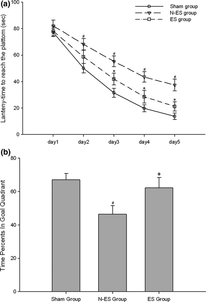
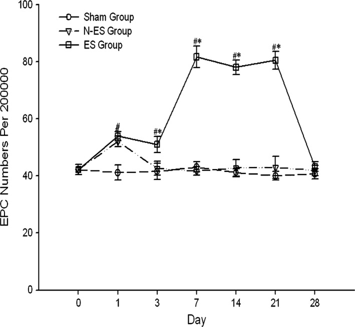
Rats were randomly divided into three groups: the Sham control group, electrical stimulation group (ES group), and No electrical stimulation control group (N-ES group). Following fluid percussion injury, the rats in the ES group received ES treatment for 3 weeks. Potent cognitive function-relevant factors, including the escape latency, time percentage in the goal quadrant, and numbers of CD34 cells, von Willebrand Factor (vWF ) vessels, and circulating endothelial progenitor cells (EPCs), were subsequently assessed using the Morris water maze (MWM) test, immunohistochemical staining, and flow cytometry.
Compared with the rats in the N-ES group, the rats in the ES group exhibited a shorter escape latency on day 3 (=.025), day 4 (=.011), and day 5 (=.003), as well as a higher time percentage in the goal quadrant (=.025) in the MWM test. After 3 weeks of ES, there were increased numbers of CD34 cells (=.008) and vWF vessels (=.000) in the hippocampus of injured brain tissue in the ES group compared with those in the N-ES group. Moreover, ES also significantly increased the number of EPCs in the peripheral blood from days 3 to 21 after TBI in the ES group (<.05).
Taken together, these findings suggest that ES may improve cognitive deficits induced by TBI, and this protective effect may be a result, in part, of enhanced angiogenesis, which may be attributed to the increased mobilization of EPCs in peripheral blood.
与创伤性脑损伤(TBI)相关的认知缺陷降低了患者的生活质量。然而,迄今为止,尚无有效的 TBI 相关认知缺陷治疗方法。在这项研究中,我们旨在确定电刺激(ES)是否改善 TBI 大鼠的认知缺陷。
大鼠随机分为三组:假手术对照组、电刺激组(ES 组)和无电刺激对照组(N-ES 组)。在液性冲击伤后,ES 组大鼠接受 ES 治疗 3 周。随后使用 Morris 水迷宫(MWM)测试、免疫组织化学染色和流式细胞术评估与认知功能相关的潜在因素,包括逃避潜伏期、目标象限时间百分比和 CD34 细胞、血管性血友病因子(vWF)血管和循环内皮祖细胞(EPCs)的数量。
与 N-ES 组大鼠相比,ES 组大鼠在 MWM 测试中第 3 天(=.025)、第 4 天(=.011)和第 5 天(=.003)的逃避潜伏期更短,目标象限的时间百分比更高(=.025)。在 ES 治疗 3 周后,ES 组大鼠海马组织损伤中 CD34 细胞的数量(=.008)和 vWF 血管的数量(=.000)均高于 N-ES 组。此外,ES 还显著增加了 TBI 后第 3 天至第 21 天外周血中 EPCs 的数量(<.05)。
综上所述,这些发现表明 ES 可能改善 TBI 引起的认知缺陷,这种保护作用部分可能是由于血管生成增强所致,这可能归因于外周血中 EPCs 的动员增加。