Ogasawara Yasushi, Kashimura Hiroshi, Aso Kenta, Saura Hiroaki
Department of Neurosurgery, Iwate Prefectural Chubu Hospital, Kitakami, Iwate, Japan.
J Neurosci Rural Pract. 2017 Oct-Dec;8(4):654-656. doi: 10.4103/jnrp.jnrp_285_17.
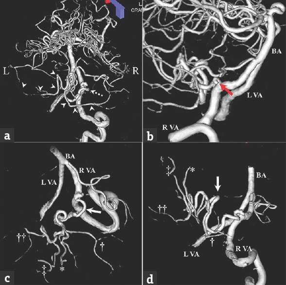
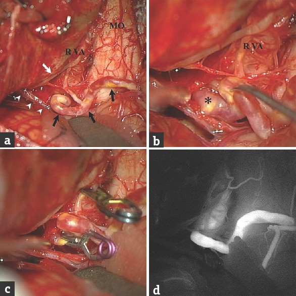
Although the anatomy of the posterior inferior cerebellar artery (PICA) is highly variable, a solitary PICA supplying both hemispheres of the cerebellum is rare. A 76-year-old woman presented with severe headache and subsequent loss of consciousness and was admitted to our hospital. Initial computed tomography showed subarachnoid hemorrhage. Three-dimensional digital subtraction angiography revealed a saccular aneurysm arising from the right vertebral artery (VA)-PICA bifurcation. The PICA branching from the right VA was enlarged, tortuous, and crossed the midline to supply both cerebellar hemispheres. This right PICA was interpreted as a bihemispheric PICA. Recognizing this variant preoperatively could help prevent complications of surgery. Careful follow-up studies are necessary in cases with bihemispheric PICA to monitor for the development of aneurysm at the junction between the bihemispheric PICA and the VA or the distal portion of the bihemispheric PICA.
尽管小脑后下动脉(PICA)的解剖结构高度变异,但由单一PICA供应小脑两个半球的情况罕见。一名76岁女性因严重头痛继而意识丧失入院。初次计算机断层扫描显示蛛网膜下腔出血。三维数字减影血管造影显示一个囊状动脉瘤起自右侧椎动脉(VA)-PICA分叉处。从右侧VA发出的PICA增粗、迂曲,并越过中线供应小脑两个半球。这条右侧PICA被判定为双半球PICA。术前识别这种变异有助于预防手术并发症。对于双半球PICA病例,有必要进行仔细的随访研究,以监测双半球PICA与VA交界处或双半球PICA远端动脉瘤的发生情况。