Rokugawa Takemi, Momosaki Sotaro, Ito Miwa, Iimori Hitoshi, Kato Yuki, Abe Kohji
Translational Research Unit, Biomarker R&D Department, Shionogi Co., Ltd, 3-1-1, Futaba-cho, Toyonaka, Osaka, 561-0825, Japan.
Department of Applied Chemistry & Analysis, Research Laboratory for Development, Shionogi & Co., Ltd, Osaka, Japan.
EJNMMI Res. 2017 Dec 6;7(1):96. doi: 10.1186/s13550-017-0345-5.
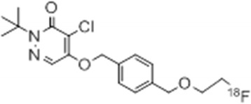
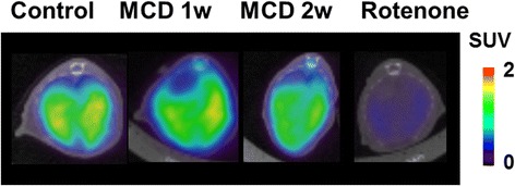
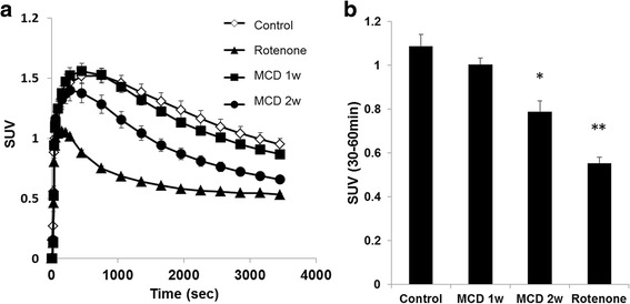
Mitochondrial dysfunction is one of the main causes of non-alcohol fatty liver disease (NAFLD). [F]-BMS-747158-02 (F-BMS) which was originally developed as a myocardial perfusion imaging agent was reported to bind mitochondrial complex-1 (MC-1). The aim of this study was to investigate the potential use of F-BMS for evaluating hepatic MC-1 activity in mice fed a methionine- and choline-deficient (MCD) diet. Male C57BL/6J mice were fed a MCD diet for up to 2 weeks. PET scans with F-BMS were performed after 1 and 2 weeks of the MCD diet. F-BMS was intravenously injected into mice, and the uptake (standardized uptake value (SUV)) in the liver was determined. The binding specificity for MC-1 was assessed by pre-administration of rotenone, a specific MC-1 inhibitor. Hepatic MC-1 activity was measured using liver homogenates generated after each positron emission tomography (PET) scan. Blood biochemistry and histopathology were also assessed.
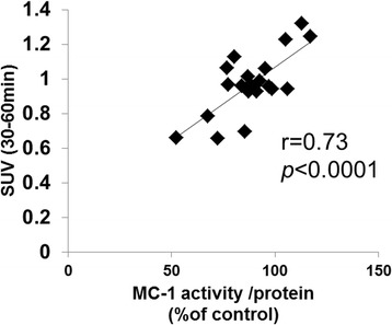
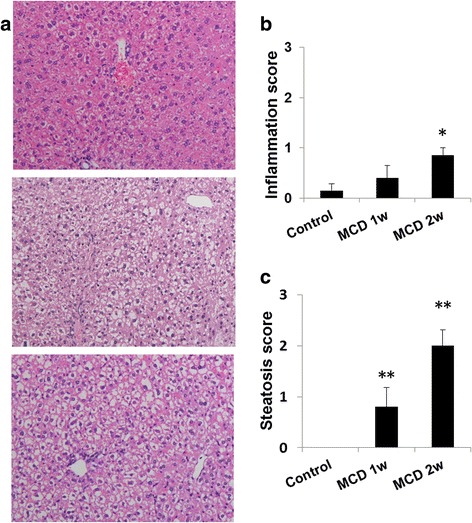
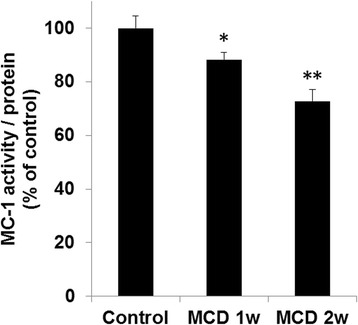
In control mice, hepatic F-BMS uptake was significantly inhibited by the pre-injection of rotenone. The uptake of F-BMS was significantly decreased after 2 weeks of the MCD diet. The SUV at 30-60 min was well correlated with hepatic MC-1 activity (r = 0.73, p < 0.05). Increases in plasma ALT and AST levels were also noted at 1 and 2 weeks. Mild hepatic steatosis with or without minimal inflammation was histopathologically observed at 1 and 2 weeks in mice liver on the MCD diet. However, inflammation was observed only at 2 weeks in mice on the MCD diet.
The present study demonstrated that F-BMS is a potential PET probe for quantitative imaging of hepatic MC-1 activity and its mitochondrial dysfunction induced by steatosis and inflammation, such as in NAFLD.
线粒体功能障碍是非酒精性脂肪性肝病(NAFLD)的主要病因之一。最初作为心肌灌注显像剂开发的[F]-BMS-747158-02(F-BMS)据报道可结合线粒体复合物I(MC-1)。本研究的目的是探讨F-BMS在评估喂食蛋氨酸和胆碱缺乏(MCD)饮食小鼠肝脏MC-1活性方面的潜在用途。雄性C57BL/6J小鼠喂食MCD饮食长达2周。在MCD饮食1周和2周后进行F-BMS的PET扫描。将F-BMS静脉注射到小鼠体内,并测定肝脏中的摄取量(标准化摄取值(SUV))。通过预先给予鱼藤酮(一种特异性MC-1抑制剂)来评估对MC-1的结合特异性。使用每次正电子发射断层扫描(PET)扫描后生成的肝脏匀浆测量肝脏MC-1活性。还评估了血液生化和组织病理学。
在对照小鼠中,预先注射鱼藤酮可显著抑制肝脏对F-BMS的摄取。MCD饮食2周后,F-BMS的摄取量显著降低。30 - 60分钟时的SUV与肝脏MC-1活性显著相关(r = 0.73,p < 0.05)。在1周和2周时还观察到血浆ALT和AST水平升高。在MCD饮食的小鼠肝脏中,在1周和2周时组织病理学观察到轻度肝脂肪变性,伴有或不伴有轻微炎症。然而,在MCD饮食的小鼠中仅在2周时观察到炎症。
本研究表明,F-BMS是一种潜在的PET探针,可用于对肝脏MC-1活性及其由脂肪变性和炎症(如在NAFLD中)诱导的线粒体功能障碍进行定量成像。