From the Department of Microbiology and Immunology (A.E.V., C.M.R.B., M.D.W.), Department of Pathology and Translational Pathobiology (J.M.G., B.H.P., A.W.O.), Department of Cell Biology and Anatomy (A.C.F.), Feist-Weiller Cancer Center (D.T.C.), and Pharmacology, Toxicology, and Neuroscience (R.L.K.), Louisiana State University Health Sciences Center, Shreveport; Department of Pharmacology, University of California San Diego, La Jolla (A.R.N.); Department of Neuroscience, Karolinska Institutet, Stockholm, Sweden (R.C.); and Division of Geriatrics and Nutritional Science, Washington University School of Medicine, St. Louis, MO (B.N.F.).
Arterioscler Thromb Vasc Biol. 2018 Feb;38(2):324-334. doi: 10.1161/ATVBAHA.117.310455. Epub 2017 Dec 7.
Macrophage proinflammatory responses induced by modified low-density lipoproteins (modLDL) contribute to atherosclerotic progression. How modLDL causes macrophages to become proinflammatory is still enigmatic. Macrophage foam cell formation induced by modLDL requires glycerolipid synthesis. Lipin-1, a key enzyme in the glycerolipid synthesis pathway, contributes to modLDL-elicited macrophage proinflammatory responses in vitro. The objective of this study was to determine whether macrophage-associated lipin-1 contributes to atherogenesis and to assess its role in modLDL-mediated signaling in macrophages.
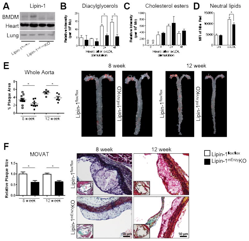
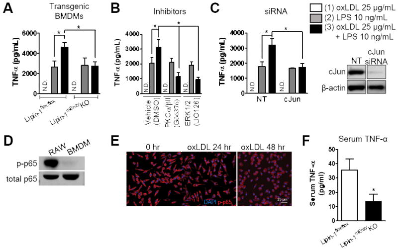
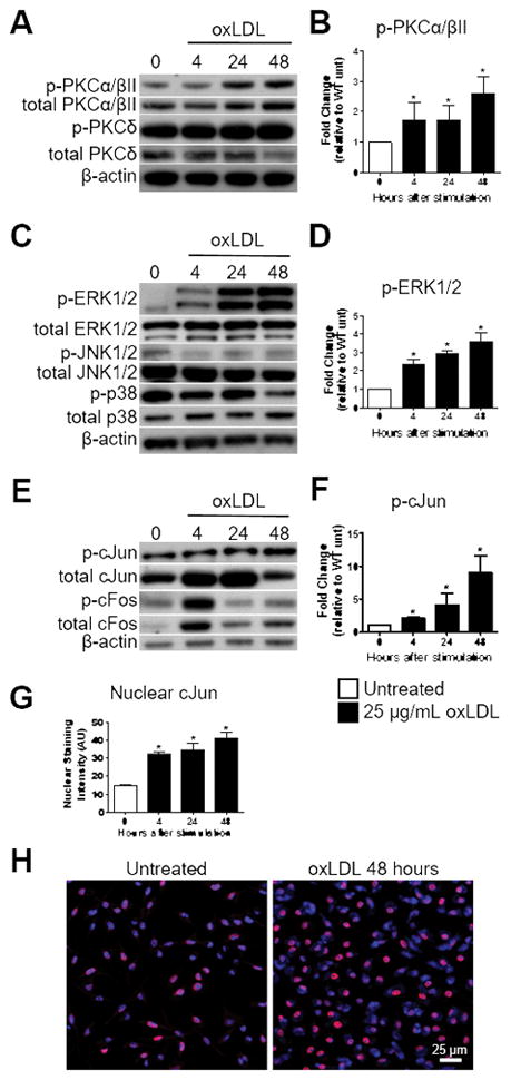
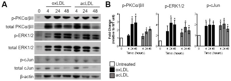
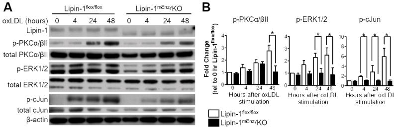
We developed mice lacking lipin-1 in myeloid-derived cells and used adeno-associated viral vector 8 expressing the gain-of-function mutation of mouse proprotein convertase subtilisin/kexin type 9 (adeno-associated viral vector 8-proprotein convertase subtilisin/kexin type 9) to induce hypercholesterolemia and plaque formation. Mice lacking myeloid-associated lipin-1 had reduced atherosclerotic burden compared with control mice despite similar plasma lipid levels. Stimulation of bone marrow-derived macrophages with modLDL activated a persistent protein kinase Cα/βII-extracellular receptor kinase1/2-jun proto-oncogene signaling cascade that contributed to macrophage proinflammatory responses that was dependent on lipin-1 enzymatic activity.
Our data demonstrate that macrophage-associated lipin-1 is atherogenic, likely through persistent activation of a protein kinase Cα/βII-extracellular receptor kinase1/2-jun proto-oncogene signaling cascade that contributes to foam cell proinflammatory responses. Taken together, these results suggest that modLDL-induced foam cell formation and modLDL-induced macrophage proinflammatory responses are not independent consequences of modLDL stimulation but rather are both directly influenced by enhanced lipid synthesis.
修饰型低密度脂蛋白(modLDL)诱导的巨噬细胞促炎反应促进动脉粥样硬化进展。modLDL 如何使巨噬细胞产生促炎作用仍不清楚。modLDL 诱导的巨噬细胞泡沫细胞形成需要甘油脂质合成。脂联素-1 是甘油脂质合成途径中的关键酶,它有助于体外 modLDL 诱导的巨噬细胞促炎反应。本研究旨在确定巨噬细胞相关脂联素-1 是否有助于动脉粥样硬化形成,并评估其在 modLDL 介导的巨噬细胞信号转导中的作用。
我们构建了骨髓细胞中缺乏脂联素-1 的小鼠,并使用表达鼠蛋白原转化酶枯草溶菌素/凝血酶 9 (腺相关病毒 8-蛋白原转化酶枯草溶菌素/凝血酶 9)的腺相关病毒 8 诱导高胆固醇血症和斑块形成。与对照小鼠相比,缺乏骨髓细胞相关脂联素-1 的小鼠尽管血浆脂质水平相似,但动脉粥样硬化负担减少。用 modLDL 刺激骨髓源性巨噬细胞激活了持续的蛋白激酶 Cα/βII-细胞外受体激酶 1/2-原癌基因 jun 信号级联反应,这有助于巨噬细胞促炎反应,而这依赖于脂联素-1 的酶活性。
我们的数据表明,巨噬细胞相关脂联素-1 是致动脉粥样硬化的,可能是通过持续激活蛋白激酶 Cα/βII-细胞外受体激酶 1/2-原癌基因 jun 信号级联反应,促进泡沫细胞的促炎反应。总之,这些结果表明,modLDL 诱导的泡沫细胞形成和 modLDL 诱导的巨噬细胞促炎反应不是 modLDL 刺激的独立后果,而是都直接受到增强的脂质合成的影响。