Vascular Biology Laboratory, Heart Institute (Incor), University of São Paulo School of Medicine, São Paulo, Brazil.
Institut für Kardiovaskuläre Physiologie, Goethe University, Frankfurt, Germany.
Sci Rep. 2017 Dec 8;7(1):17262. doi: 10.1038/s41598-017-16947-5.
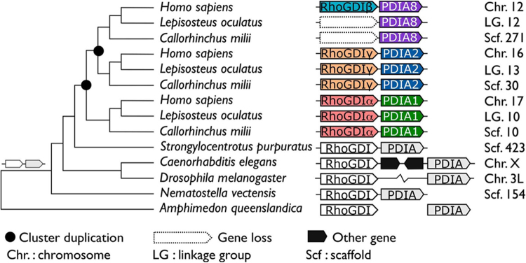
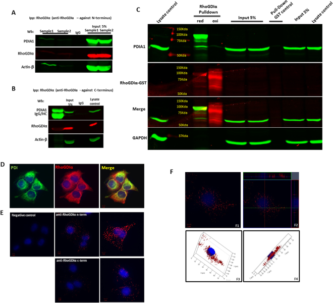
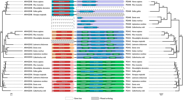
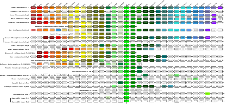
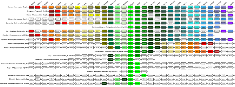
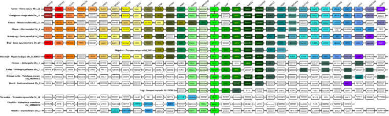
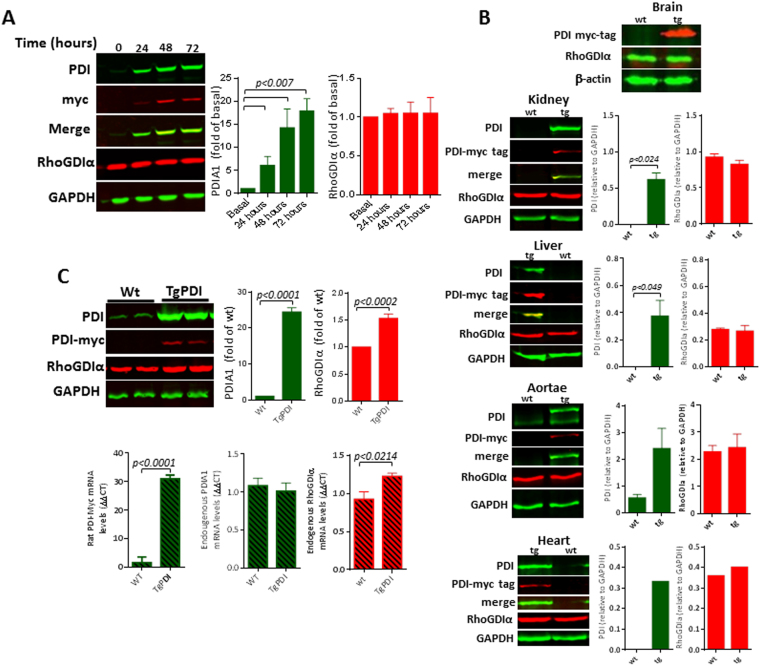
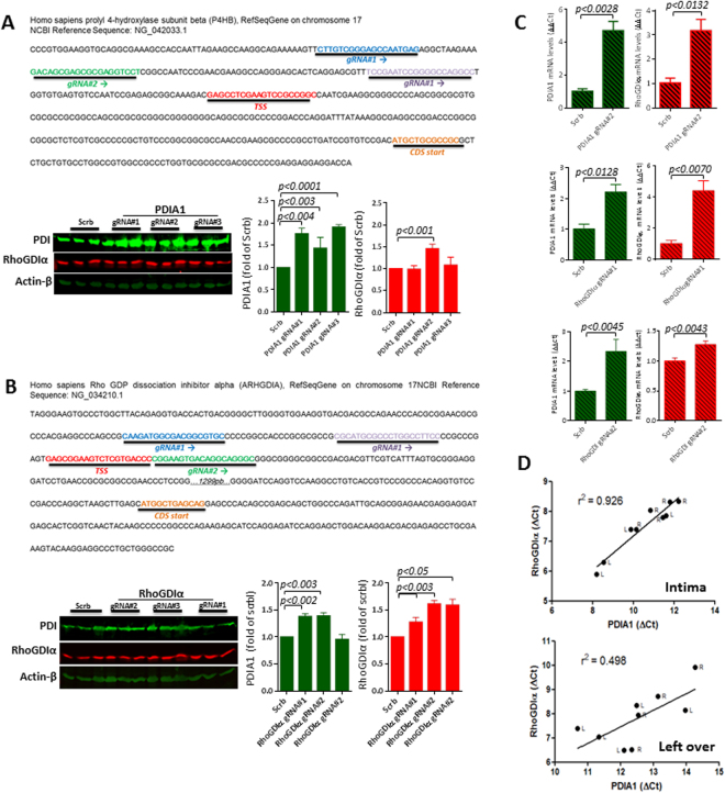
Protein disulfide isomerases (PDIs) support endoplasmic reticulum redox protein folding and cell-surface thiol-redox control of thrombosis and vascular remodeling. The family prototype PDIA1 regulates NADPH oxidase signaling and cytoskeleton organization, however the related underlying mechanisms are unclear. Here we show that genes encoding human PDIA1 and its two paralogs PDIA8 and PDIA2 are each flanked by genes encoding Rho guanine-dissociation inhibitors (GDI), known regulators of RhoGTPases/cytoskeleton. Evolutionary histories of these three microsyntenic regions reveal their emergence by two successive duplication events of a primordial gene pair in the last common vertebrate ancestor. The arrangement, however, is substantially older, detectable in echinoderms, nematodes, and cnidarians. Thus, PDI/RhoGDI pairing in the same transcription orientation emerged early in animal evolution and has been largely maintained. PDI/RhoGDI pairs are embedded into conserved genomic regions displaying common cis-regulatory elements. Analysis of gene expression datasets supports evidence for PDI/RhoGDI coexpression in developmental/inflammatory contexts. PDIA1/RhoGDIα were co-induced in endothelial cells upon CRISP-R-promoted transcription activation of each pair component, and also in mouse arterial intima during flow-induced remodeling. We provide evidence for physical interaction between both proteins. These data support strong functional links between PDI and RhoGDI families, which likely maintained PDI/RhoGDI microsynteny along > 800-million years of evolution.
蛋白质二硫键异构酶(PDI)支持内质网氧化还原蛋白折叠和细胞表面硫醇氧化还原对血栓形成和血管重塑的控制。家族原型 PDIA1 调节 NADPH 氧化酶信号和细胞骨架组织,但其相关潜在机制尚不清楚。在这里,我们表明,编码人 PDIA1 及其两个同源物 PDIA8 和 PDIA2 的基因分别被编码 Rho 鸟嘌呤解离抑制剂(GDI)的基因所包围,RhoGTPases/细胞骨架的已知调节剂。这三个微同源区域的进化历史揭示了它们是由原始基因对在最后一个共同脊椎动物祖先中的两次连续复制事件产生的。然而,这种排列方式要古老得多,在棘皮动物、线虫和刺胞动物中都可以检测到。因此,PDI/RhoGDI 在同一转录方向上的配对在动物进化的早期就出现了,并在很大程度上得到了维持。PDI/RhoGDI 对嵌入到具有共同顺式调控元件的保守基因组区域中。基因表达数据集的分析支持在发育/炎症背景下 PDI/RhoGDI 共表达的证据。在 CRISP-R 促进的每个配对成分的转录激活后,内皮细胞中 PDIA1/RhoGDIα 被共同诱导,在血流诱导的重塑过程中,小鼠动脉内膜也是如此。我们提供了这两种蛋白质之间物理相互作用的证据。这些数据支持 PDI 和 RhoGDI 家族之间的强烈功能联系,这可能沿着超过 8 亿年的进化过程维持了 PDI/RhoGDI 的微同源性。