Picower Institute for Learning and Memory, Cambridge, MA 02139.
Department of Brain and Cognitive Sciences, Massachusetts Institute of Technology, Cambridge, MA 02139.
eNeuro. 2017 Dec 15;4(6). doi: 10.1523/ENEURO.0163-15.2017. eCollection 2017 Nov-Dec.
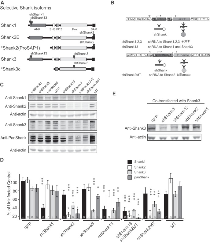
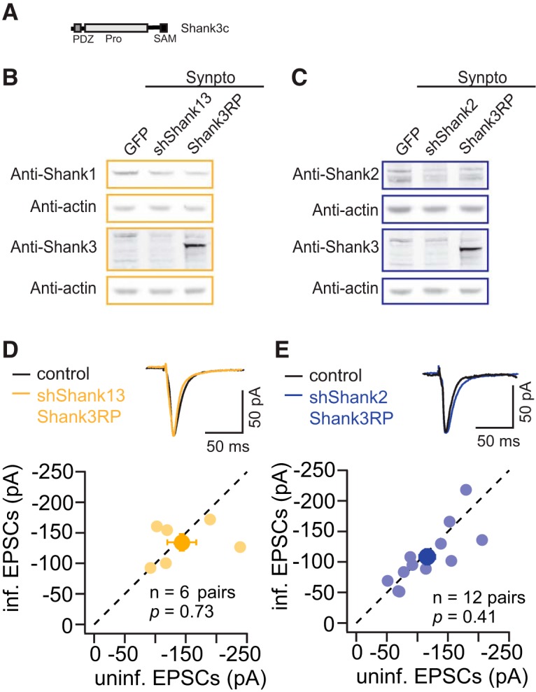
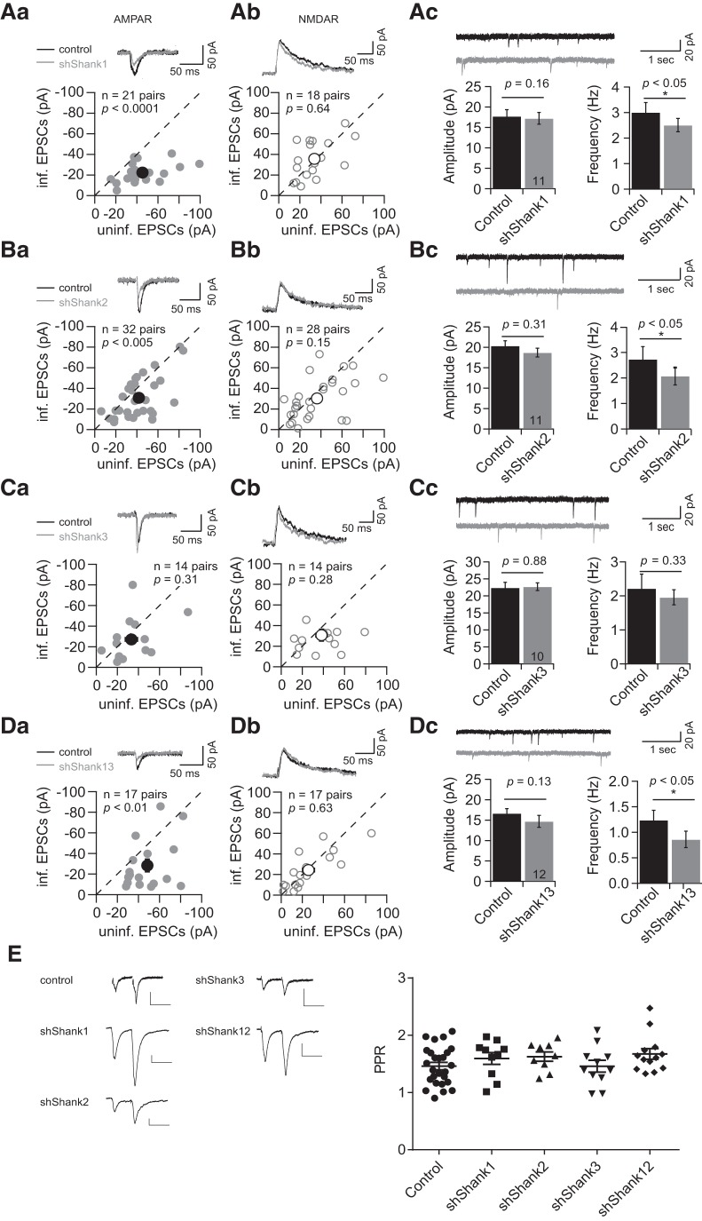
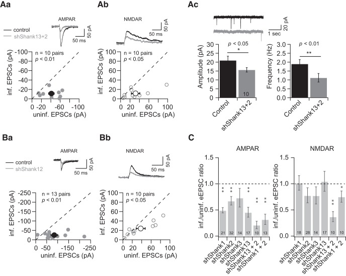
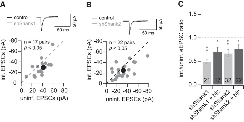
Shank proteins, one of the principal scaffolds in the postsynaptic density (PSD) of the glutamatergic synapses, have been associated with autism spectrum disorders and neuropsychiatric diseases. However, it is not known whether different Shank family proteins have distinct functions in regulating synaptic transmission, and how they differ from other scaffold proteins in this aspect. Here, we investigate the role of Shanks in regulating glutamatergic synaptic transmission at rat hippocampal SC-CA1 synapses, using lentivirus-mediated knockdown and molecular replacement combined with dual whole-cell patch clamp in hippocampal slice culture. In line with previous findings regarding PSD-MAGUK scaffold manipulation, we found that loss of scaffold proteins via knockdown of Shank1 or Shank2, but not Shank3, led to a reduction of the number but not the unitary response of AMPAR-containing synapses. Only when both Shank1 and Shank2 were knocked down, were both the number and the unitary response of active synapses reduced. This reduction was accompanied by a decrease in NMDAR-mediated synaptic response, indicating more profound deficits in synaptic transmission. Molecular replacement with Shank2 and Shank3c rescued the synaptic transmission to the basal level, and the intact sterile α-motif (SAM) of Shank proteins is required for maintaining glutamatergic synaptic transmission. We also found that altered neural activity did not influence the effect of Shank1 or Shank2 knockdown on AMPAR synaptic transmission, in direct contrast to the activity dependence of the effect of PSD-95 knockdown, revealing differential interaction between activity-dependent signaling and scaffold protein families in regulating synaptic AMPAR function.
Shank 蛋白是谷氨酸能突触后密度(PSD)的主要支架之一,与自闭症谱系障碍和神经精神疾病有关。然而,目前尚不清楚不同的 Shank 家族蛋白在调节突触传递方面是否具有不同的功能,以及它们在这方面与其他支架蛋白有何不同。在这里,我们使用慢病毒介导的敲低和分子置换结合海马脑片培养中的双全细胞膜片钳技术,研究了 Shank 在调节大鼠海马 SC-CA1 突触谷氨酸能突触传递中的作用。与先前关于 PSD-MAGUK 支架操作的发现一致,我们发现通过敲低 Shank1 或 Shank2 而不是 Shank3 损失支架蛋白会导致 AMPAR 包含的突触数量减少,但单位反应不变。只有当 Shank1 和 Shank2 都被敲低时,活跃突触的数量和单位反应才会减少。这种减少伴随着 NMDAR 介导的突触反应减少,表明突触传递有更严重的缺陷。用 Shank2 和 Shank3c 进行分子置换可将突触传递恢复到基础水平,Shank 蛋白的完整无菌 α 基序(SAM)对于维持谷氨酸能突触传递是必需的。我们还发现,改变神经活动不会影响 Shank1 或 Shank2 敲低对 AMPAR 突触传递的影响,这与 PSD-95 敲低的活动依赖性效应形成直接对比,揭示了在调节突触 AMPAR 功能方面,活性依赖性信号和支架蛋白家族之间的差异相互作用。