Department of Medical Sciences, University of Turin, Turin, Italy.
Clinical Biochemistry Laboratory, "Città della Salute e della Scienza" Hospital of Turin, Turin, Italy.
Nutr Diabetes. 2017 Dec 18;7(12):303. doi: 10.1038/s41387-017-0010-0.
Timing of food intake impacts on metabolic diseases. Few data are available about post-meal changes in epinephrine (E), norepinephrine (NE), and acylated ghrelin (AG) at different times of the day.
This randomized cross-over trial investigated E/NE/AG concentrations after identical meals consumed at 0800 or 2000 hours in 20 healthy volunteers, by standardizing diet, exercise, duration of fast, and resting. Participants randomly received the test meal at 0800 or 2000 hours, and vice versa after 1 week. Blood samples were collected before and up to 180-min post-meal, every 30 min, with participants supine, motionless, but awake.
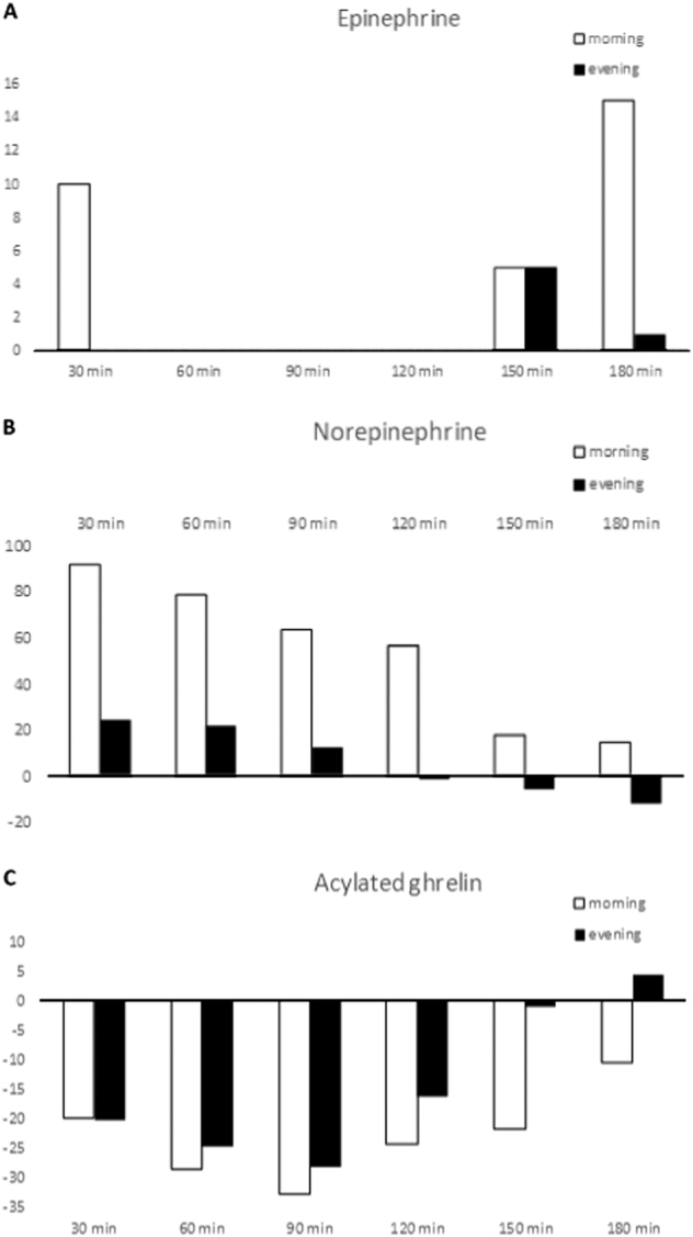
Median E levels increased at 30-60 min, then declined and rose again at 150 min; values at 60 min (19.0 vs. 15.0 ng/l, p = 0.03) and 180 min (25.0 vs. 11.0 ng/l, p < 0.001) were higher after the morning meals. NE rose at 30-60 min and then progressively declined; median values at 60 min (235.3 vs. 206.3 ng/l, p = 0.02) and 120 min (208.8 vs. 142.0 ng/l, p = 0.04) increased more after morning meals. AG progressively declined to increase again at 90 min after meal; median AG area-under-the-curve (AUC) values were lower at morning (7206.8 vs. 8828.3 pg/mL×h). AG-AUC was inversely associated with diet-induced thermogenesis (β = -121.6; 95% CI -201.0 to 42.2; p = 0.009 for each unit increase), while log NE-AUC was inversely associated with log-triglyceride AUC (β = -0.57; 95% CI -0.98 to 0.16; p = 0.015) in a multiple regression model, after multiple adjustments.
In conclusion, E/NE concentrations were higher after the morning meal, while AG showed an opposite behavior. These data, although requiring confirmation in larger samples, suggest an adjunctive possible mechanism explaining the unfavorable effects of evening eating on metabolic risk.
饮食时间会影响代谢疾病。关于一天中不同时间餐后肾上腺素(E)、去甲肾上腺素(NE)和酰化 ghrelin(AG)的变化,数据很少。
本随机交叉试验研究了 20 名健康志愿者在 0800 或 2000 时摄入相同的餐后,E/NE/AG 浓度的变化,通过标准化饮食、运动、禁食时间和休息。参与者随机在 0800 或 2000 时接受测试餐,1 周后反之亦然。在餐后 0 至 180 分钟内,每 30 分钟采集一次血液样本,参与者保持仰卧位、静止但清醒。
E 水平在 30-60 分钟时升高,然后下降并在 150 分钟时再次升高;60 分钟(19.0 与 15.0ng/l,p=0.03)和 180 分钟(25.0 与 11.0ng/l,p<0.001)时的数值更高进食后的早餐。NE 在 30-60 分钟时升高,然后逐渐下降;60 分钟(235.3 与 206.3ng/l,p=0.02)和 120 分钟(208.8 与 142.0ng/l,p=0.04)时的中位值升高更多在早餐后。AG 逐渐下降,90 分钟后再次增加;早餐时 AG 曲线下面积(AUC)的中位数值较低(7206.8 与 8828.3pg/mL×h)。AG-AUC 与饮食诱导的产热呈负相关(β=-121.6;95%CI-201.0 至 42.2;每增加一个单位,p=0.009),而在多元回归模型中,logNE-AUC 与 log-甘油三酯 AUC 呈负相关(β=-0.57;95%CI-0.98 至 0.16;p=0.015),经过多次调整。
总之,早餐后 E/NE 浓度升高,而 AG 则表现出相反的行为。尽管这些数据需要在更大的样本中进行验证,但它们表明了一种补充的可能机制,可以解释晚上进食对代谢风险的不利影响。