González-Arnay Emilio, González-Gómez Miriam, Meyer Gundela
Unit of Pathology, Department of Basic Medical Science, Faculty of Medicine, University of La Laguna, San Cristóbal de La Laguna, Spain.
Unit of Anatomy, Department of Basic Medical Science, Faculty of Medicine, University of La Laguna, San Cristóbal de La Laguna, Spain.
Front Neuroanat. 2017 Dec 5;11:111. doi: 10.3389/fnana.2017.00111. eCollection 2017.
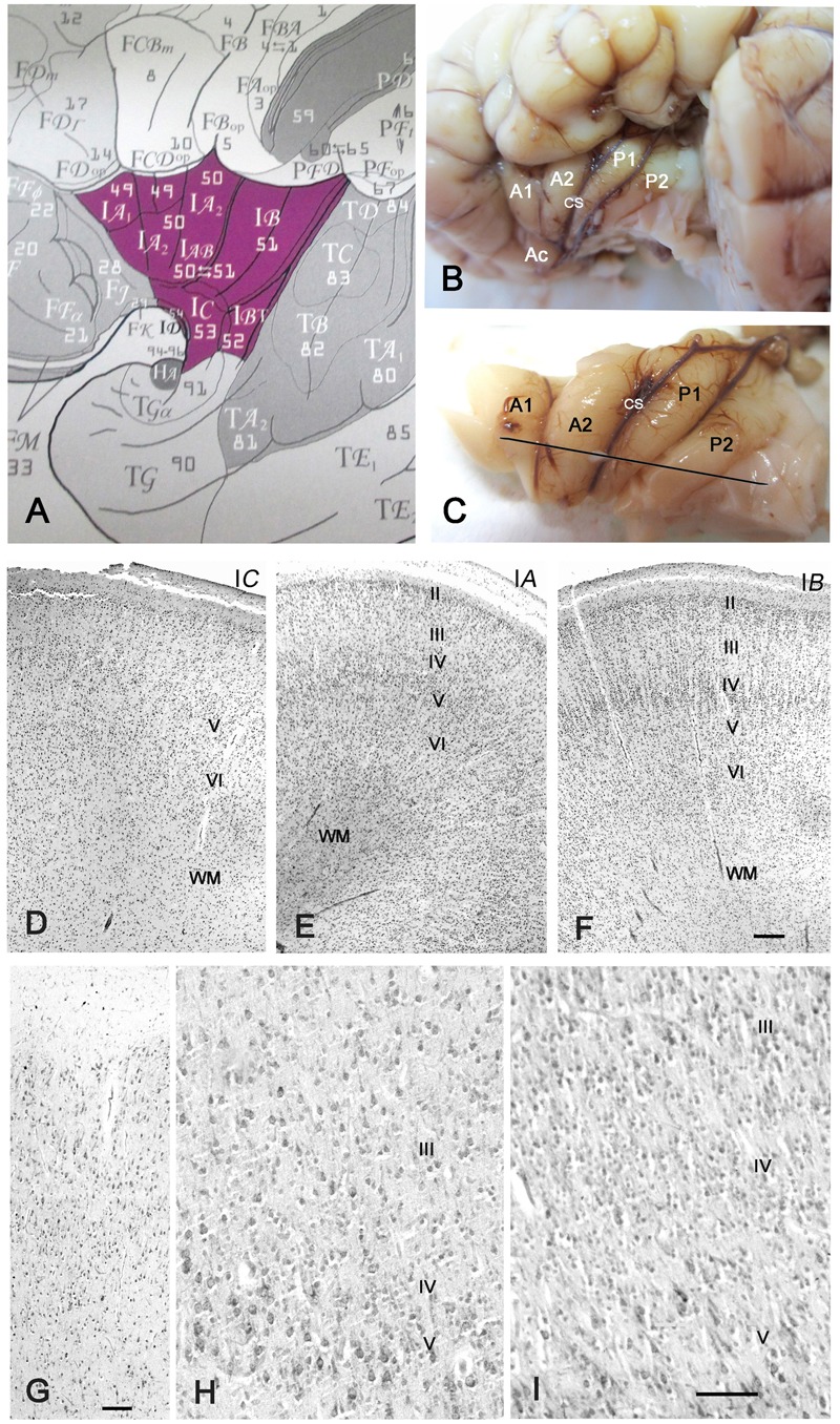
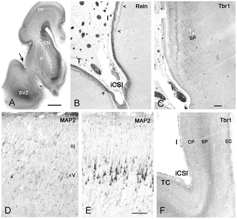
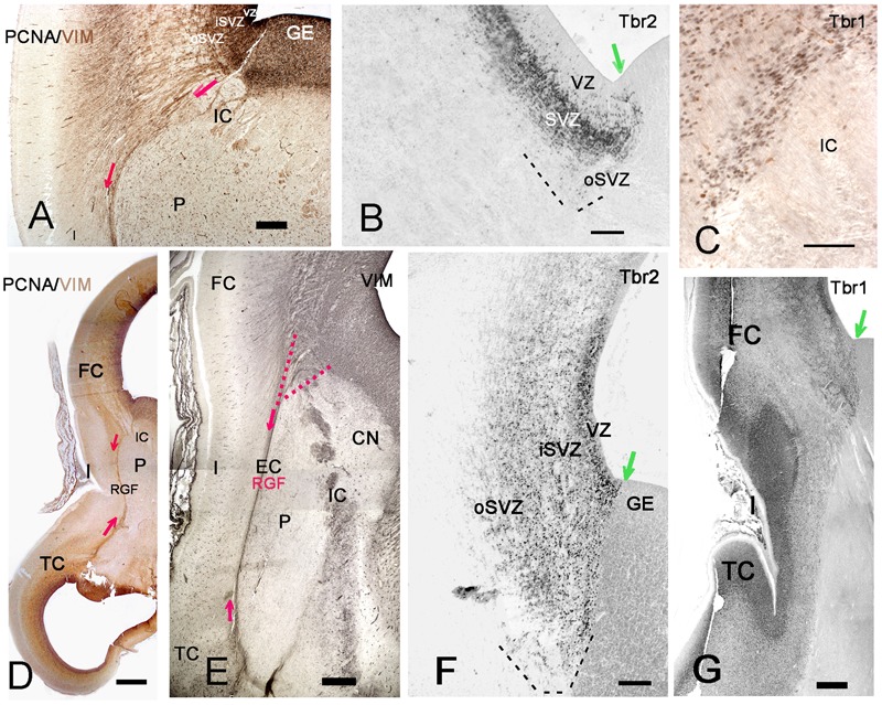
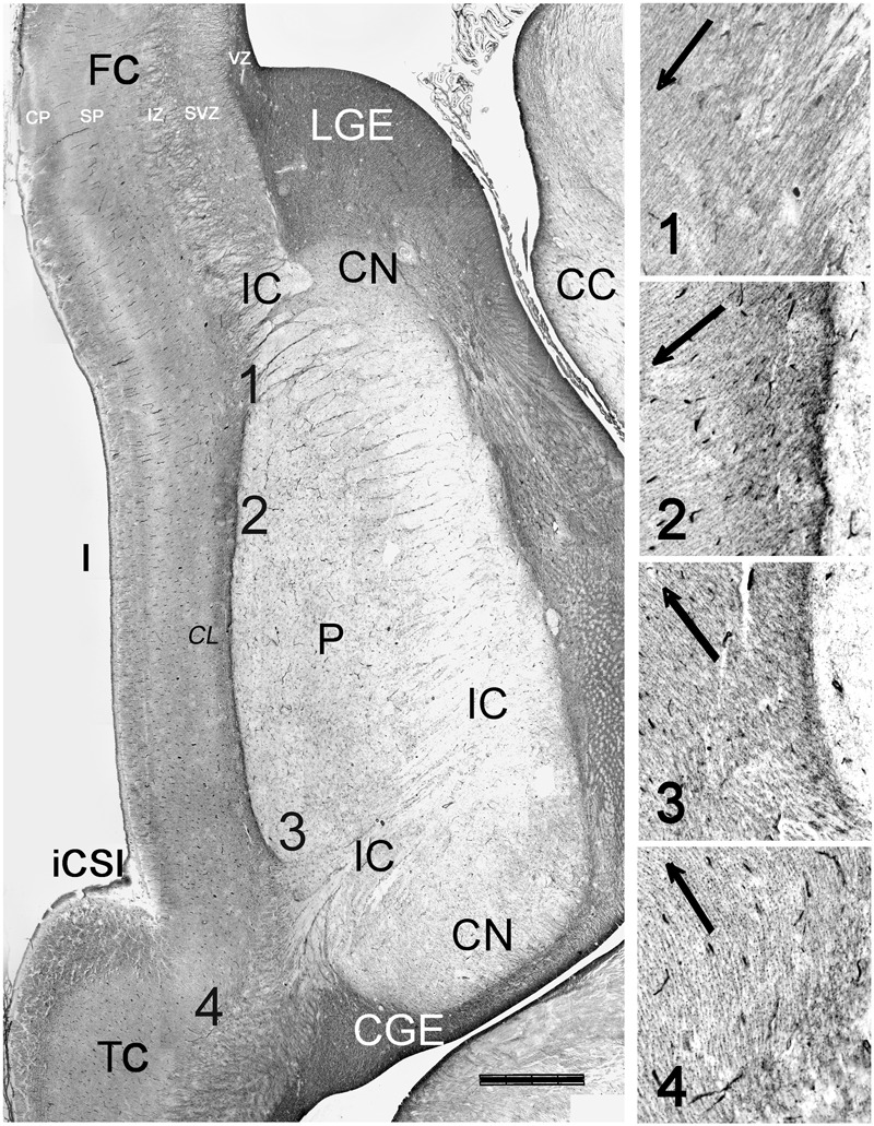
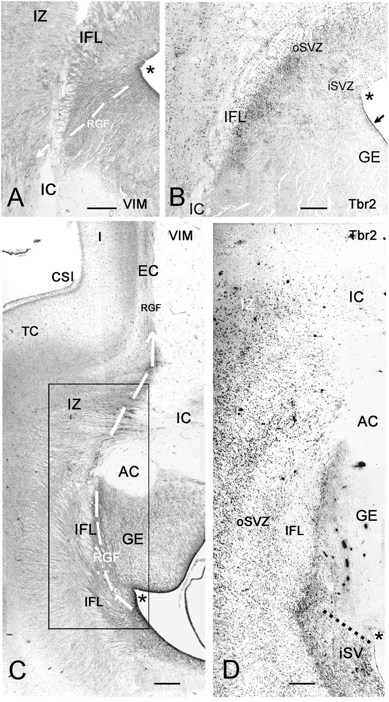
The human insular lobe, in the depth of the Sylvian fissure, displays three main cytoarchitectonic divisions defined by the differentiation of granular layers II and IV. These comprise a rostro-ventral agranular area, an intermediate dysgranular area, and a dorso-caudal granular area. Immunohistochemistry in human embryos and fetuses using antibodies against PCNA, Vimentin, Nestin, Tbr1, and Tb2 reveals that the insular cortex is unique in that it develops far away from the ventricular zone (VZ), with most of its principal neurons deriving from the subventricular zone (SVZ) of the pallial-subpallial boundary (PSB). In human embryos (Carnegie stage 16/17), the rostro-ventral insula is the first cortical region to develop; its Tbr1+ neurons migrate from the PSB along the lateral cortical stream. From 10 gestational weeks (GW) onward, lateral ventricle, ganglionic eminences, and PSB grow forming a C-shaped curvature. The SVZ of the PSB gives rise to a distinct radial glia fiber fascicle (RGF), which courses lateral to the putamen in the external capsule. In the RGF, four components can be established: PF, descending from the prefrontal PSB to the anterior insula; FP, descending from the fronto-parietal PSB toward the intermediate insula; PT, coursing from the PSB near the parieto-temporal junction to the posterior insula, and T, ascending from the temporal PSB and merging with components FP and PT. The RGF fans out at different dorso-ventral and rostro-caudal levels of the insula, with descending fibers predominating over ascending ones. The RGF guides migrating principal neurons toward the future agranular, dysgranular, and granular insular areas, which show an adult-like definition at 32 GW. Despite the narrow subplate, and the absence of an intermediate zone except in the caudal insula, most insular subdivisions develop into a 6-layered isocortex, possibly due to the well developed outer SVZ at the PSB, which is particularly prominent at the level of the dorso-caudal insula. The small size of the initial PSB sector may, however, determine the limited surface expansion of the insula, which is in contrast to the exuberant growth of the opercula deriving from the adjacent frontal-parietal and temporal VZ/SVZ.
人类脑岛叶位于外侧裂深处,根据颗粒层II和IV的分化显示出三个主要的细胞构筑分区。这些分区包括一个嘴侧腹侧无颗粒区、一个中间颗粒减少区和一个背侧尾侧颗粒区。使用抗PCNA、波形蛋白、巢蛋白、Tbr1和Tb2抗体对人类胚胎和胎儿进行免疫组织化学分析表明,脑岛皮质的独特之处在于它远离脑室区(VZ)发育,其大多数主要神经元源自皮质-皮质下边界(PSB)的室下区(SVZ)。在人类胚胎(卡内基分期16/17)中,嘴侧腹侧脑岛是第一个发育的皮质区域;其Tbr1+神经元从PSB沿着外侧皮质流迁移。从妊娠10周(GW)起,侧脑室、神经节隆起和PSB生长形成C形弯曲。PSB的SVZ产生一条独特的放射状胶质纤维束(RGF),它在外囊内沿着壳核外侧走行。在RGF中,可以确定四个成分:PF,从额叶PSB向下延伸至前脑岛;FP,从额顶叶PSB向下延伸至中间脑岛;PT,从靠近顶颞交界处的PSB走向后脑岛,以及T,从颞叶PSB向上延伸并与成分FP和PT汇合。RGF在脑岛不同的背腹侧和嘴尾侧水平呈扇形散开,下行纤维多于上行纤维。RGF引导迁移的主要神经元朝向未来的无颗粒、颗粒减少和颗粒状脑岛区域,这些区域在32GW时呈现出类似成人的结构。尽管亚板狭窄,且除了尾侧脑岛外没有中间区,但大多数脑岛亚区发育成6层的同型皮质,这可能是由于PSB处发育良好的外侧SVZ,在背侧尾侧脑岛水平尤为突出。然而,初始PSB区域的小尺寸可能决定了脑岛有限的表面扩展,这与源自相邻额顶叶和颞叶VZ/SVZ的脑盖的旺盛生长形成对比。