Wang Chuan, Li Lin, Liu Zhicheng
Department of Biomechanics and Rehabilitation Engineering, School of Biomedical Engineering, Capital Medical University, No.10 Xitoutiao, You An Men, Beijing, 100069, People's Republic of China.
Beijing Key Laboratory of Fundamental Research on Biomechanics in Clinical Application, Capital Medical University, Beijing, 100069, China.
BMC Ophthalmol. 2017 Dec 29;17(1):268. doi: 10.1186/s12886-017-0662-5.
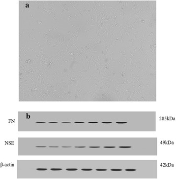
Trabecular meshwork (TM) plays an important role in maintaining normal intraocular pressure (IOP). Studies have shown that glaucomatous TM tissues are stiffer than those of normal tissue. The high expression of fibronectin protein (FN) and adaptor protein (LNK) may be related to high resistance to aqueous humor outflow as well as high IOP. Our concern is what factors lead to the variation of the stiffness of trabecular tissue/cells.
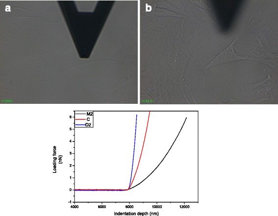
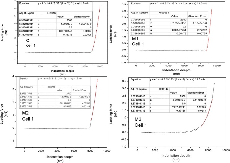
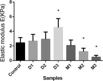
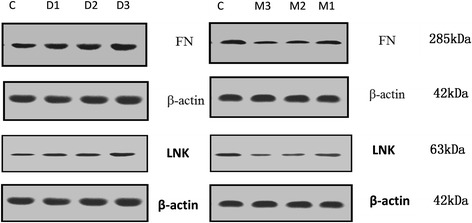
Atomic force microscope (AFM) and Western blot (WB) analysis were applied to test TM cells of rats cultured with different concentrations of dexamethasone (DEX) and mifepristone (MIF). Rat TM cells were randomly divided into 7 groups, marked as D1, D2, D3 and M1, M2 M3 for different concentrations of DEX and MIF, respectively, and C for blank control.
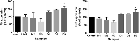
The elastic modulus of the treated cells were 2.67 ± 0.914 KPa, 2.92 ± 0.986 KPa, 4.52 ± 1.22 KPa for D1, D2, D3, 2.06 ± 0.745 KPa, 1.23 ± 0.462 KPa, 0.467 ± 0.275 KPa for M1, M2, M3, and 2.43 ± 0.713 KPa for C group, respectively. Expressions of FN and LNK increase (decrease) with the increase of the concentrations of DEX (MIF).
We focus on the relationship between the stiffness and the expressions of FN and LNK of rat TM cells. We analyzed the correlation between cell stiffness and FN, LNK expression, discussed the relationship between cell stiffness and aqueous humor outflow resistance.
The changes of TM cell stiffness and the expressions of FN and LNK are positively correlated.
小梁网(TM)在维持正常眼压(IOP)中起重要作用。研究表明,青光眼患者的小梁网组织比正常组织更硬。纤连蛋白(FN)和衔接蛋白(LNK)的高表达可能与房水流出阻力增加以及高眼压有关。我们关注的是哪些因素导致小梁组织/细胞硬度的变化。
应用原子力显微镜(AFM)和蛋白质免疫印迹法(WB)分析用不同浓度地塞米松(DEX)和米非司酮(MIF)培养的大鼠小梁网细胞。将大鼠小梁网细胞随机分为7组,分别标记为D1、D2、D3和M1、M2、M3,分别代表不同浓度的DEX和MIF,C组为空白对照。
D1、D2、D3组处理后细胞的弹性模量分别为2.67±0.914千帕、2.92±0.986千帕、4.52±1.22千帕,M1、M2、M3组分别为2.06±0.745千帕、1.23±0.462千帕、0.467±0.275千帕,C组为2.43±0.713千帕。FN和LNK的表达随DEX(MIF)浓度的增加而增加(减少)。
我们关注大鼠小梁网细胞硬度与FN和LNK表达之间的关系。分析了细胞硬度与FN、LNK表达的相关性,探讨了细胞硬度与房水流出阻力的关系。
小梁网细胞硬度变化与FN和LNK表达呈正相关。