Diagnostic Cardiovascular Imaging Laboratory, Department of Radiological Sciences, David Geffen School of Medicine at UCLA, Los Angeles, California, USA.
Division of Cardiology, David Geffen School of Medicine at UCLA and VA Greater Los Angeles Healthcare System, Los Angeles, California, USA.
J Cardiovasc Magn Reson. 2017 Dec 28;19(1):106. doi: 10.1186/s12968-017-0422-y.
Bright-blood and black-blood cardiovascular magnetic resonance (CMR) techniques are frequently employed together during a clinical exam because of their complementary features. While valuable, existing black-blood CMR approaches are flow dependent and prone to failure. We aim to assess the effectiveness and reliability of ferumoxytol enhanced (FE) Half-Fourier Single-shot Turbo Spin-echo (HASTE) imaging without magnetization preparation pulses to yield uniform intra-luminal blood signal suppression by comparing FE-HASTE with pre-ferumoxytol HASTE imaging.
This study was IRB-approved and HIPAA compliant. Consecutive patients who were referred for FE-CMR between June 2013 and February 2017 were enrolled. Qualitative image scores reflecting the degree and reliability of blood signal suppression were based on a 3-point Likert scale, with 3 reflecting perfect suppression. For quantitative evaluation, homogeneity indices (defined as standard deviation of the left atrial signal intensity) and signal-to-noise ratios (SNR) for vascular lumens and cardiac chambers were measured.
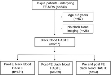
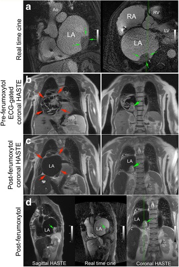
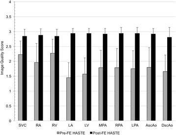
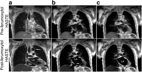
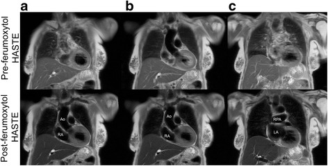
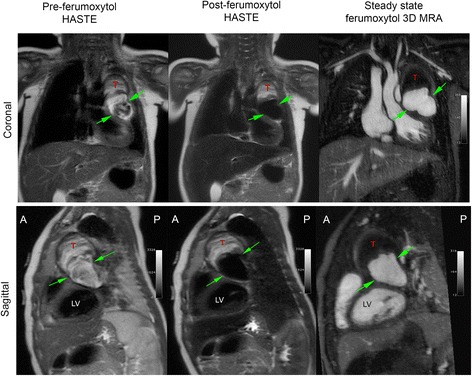
Of the 340 unique patients who underwent FE-CMR, HASTE was performed in 257. Ninety-three patients had both pre-ferumoxytol HASTE and FE-HASTE, and were included in this analysis. Qualitative image scores reflecting the degree and reliability of blood signal suppression were significantly higher for FE-HASTE images (2.9 [IQR 2.8-3.0] vs 1.8 [IQR 1.6-2.1], p < 0.001). Inter-reader agreement was moderate (k = 0.50, 95% CI 0.45-0.55). Blood signal suppression was more complete on FE-HASTE images than on pre-ferumoxytol HASTE, as indicated by lower mean homogeneity indices (24.5 [IQR 18.0-32.8] vs 108.0 [IQR 65.0-170.4], p < 0.001) and lower blood pool SNR for all regions (5.6 [IQR 3.2-10.0] vs 21.5 [IQR 12.5-39.4], p < 0.001).
FE-HASTE black-blood imaging offers an effective, reliable, and simple approach for flow independent blood signal suppression. The technique holds promise as a fast and routine complement to bright-blood cardiovascular imaging with ferumoxytol.
在临床检查中,由于其互补特性,经常同时使用亮血和黑血心血管磁共振(CMR)技术。虽然这些方法很有价值,但现有的黑血 CMR 方法依赖于血流,并且容易出现故障。我们旨在评估无磁化准备脉冲的铁氧体增强(FE)半傅立叶单 shot 涡轮自旋回波(HASTE)成像的有效性和可靠性,通过比较 FE-HASTE 与预铁氧体 HASTE 成像,以产生均匀的管腔内血液信号抑制。
本研究获得了机构审查委员会的批准和符合 HIPAA 标准。连续入组 2013 年 6 月至 2017 年 2 月期间因 FE-CMR 而转介的患者。基于 3 分李克特量表评估反映血液信号抑制程度和可靠性的定性图像评分,3 分表示完全抑制。进行定量评估时,测量左心房信号强度的标准差(定义为均匀性指数)和血管腔和心腔的信噪比(SNR)。
在 340 例接受 FE-CMR 的患者中,257 例行 HASTE 检查。93 例患者均进行了预铁氧体 HASTE 和 FE-HASTE 检查,并纳入本分析。FE-HASTE 图像的血液信号抑制程度和可靠性的定性图像评分明显更高(2.9[IQR 2.8-3.0] 比 1.8[IQR 1.6-2.1],p<0.001)。两位读者之间的一致性为中度(k=0.50,95%CI 0.45-0.55)。FE-HASTE 图像上的血液信号抑制更完全,表明平均均匀性指数较低(24.5[IQR 18.0-32.8] 比 108.0[IQR 65.0-170.4],p<0.001),所有区域的血池 SNR 均较低(5.6[IQR 3.2-10.0] 比 21.5[IQR 12.5-39.4],p<0.001)。
FE-HASTE 黑血成像提供了一种有效、可靠且简单的血流独立血液信号抑制方法。该技术有望成为铁氧体增强后的快速常规亮血心血管成像的补充方法。