Budde Matthew D, Skinner Nathan P, Muftuler L Tugan, Schmit Brian D, Kurpad Shekar N
Department of Neurosurgery, Medical College of Wisconsin, Milwaukee, WI, United States.
Medical Scientist Training Program, Medical College of Wisconsin, Milwaukee, WI, United States.
Front Neurosci. 2017 Dec 19;11:706. doi: 10.3389/fnins.2017.00706. eCollection 2017.
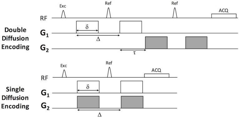
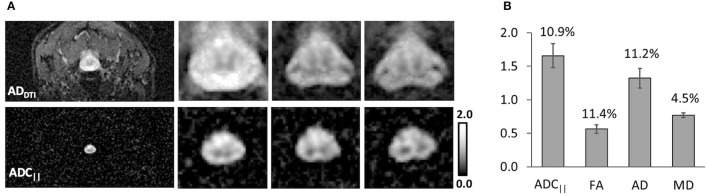
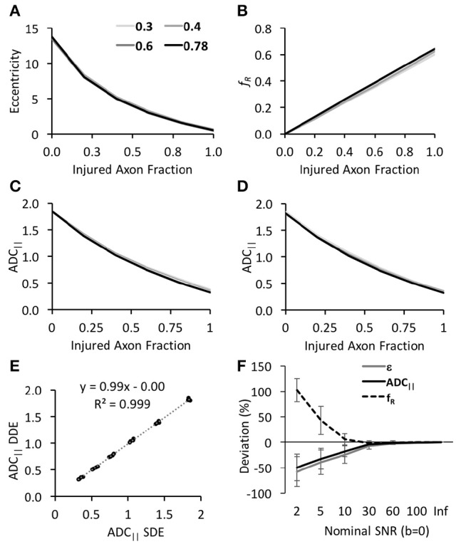
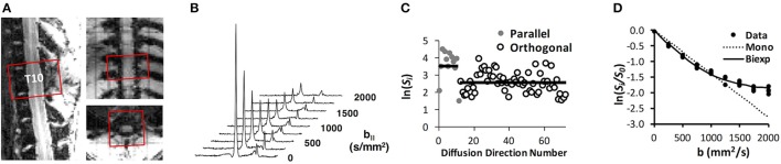
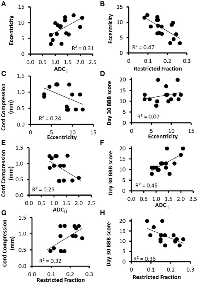
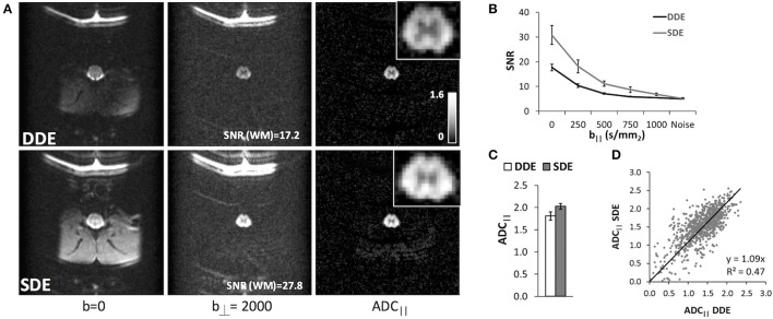
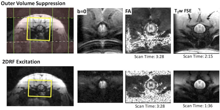
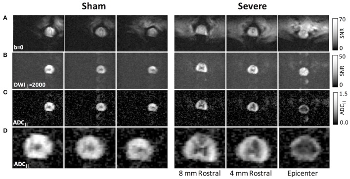
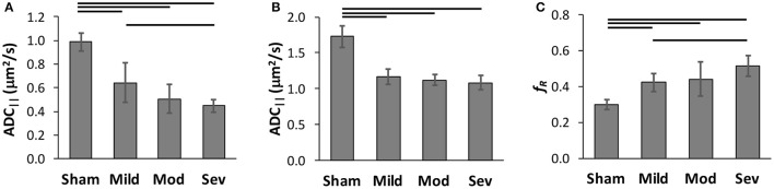
Diffusion tensor imaging (DTI) is a promising biomarker of spinal cord injury (SCI). In the acute aftermath, DTI in SCI animal models consistently demonstrates high sensitivity and prognostic performance, yet translation of DTI to acute human SCI has been limited. In addition to technical challenges, interpretation of the resulting metrics is ambiguous, with contributions in the acute setting from both axonal injury and edema. Novel diffusion MRI acquisition strategies such as double diffusion encoding (DDE) have recently enabled detection of features not available with DTI or similar methods. In this work, we perform a systematic optimization of DDE using simulations and an rat model of SCI and subsequently implement the protocol to the healthy human spinal cord. First, two complementary DDE approaches were evaluated using an orientationally invariant or a filter-probe diffusion encoding approach. While the two methods were similar in their ability to detect acute SCI, the filter-probe DDE approach had greater predictive power for functional outcomes. Next, the filter-probe DDE was compared to an analogous single diffusion encoding (SDE) approach, with the results indicating that in the spinal cord, SDE provides similar contrast with improved signal to noise. In the SCI rat model, the filter-probe SDE scheme was coupled with a reduced field of view (rFOV) excitation, and the results demonstrate high quality maps of the spinal cord without contamination from edema and cerebrospinal fluid, thereby providing high sensitivity to injury severity. The optimized protocol was demonstrated in the healthy human spinal cord using the commercially-available diffusion MRI sequence with modifications only to the diffusion encoding directions. Maps of axial diffusivity devoid of CSF partial volume effects were obtained in a clinically feasible imaging time with a straightforward analysis and variability comparable to axial diffusivity derived from DTI. Overall, the results and optimizations describe a protocol that mitigates several difficulties with DTI of the spinal cord. Detection of acute axonal damage in the injured or diseased spinal cord will benefit the optimized filter-probe diffusion MRI protocol outlined here.
扩散张量成像(DTI)是一种很有前景的脊髓损伤(SCI)生物标志物。在急性损伤后,SCI动物模型中的DTI始终显示出高敏感性和预后性能,但DTI在急性人类SCI中的应用却很有限。除了技术挑战外,对所得指标的解释也不明确,因为在急性情况下,轴突损伤和水肿都会产生影响。新型扩散磁共振成像采集策略,如双扩散编码(DDE),最近使得检测DTI或类似方法无法获得的特征成为可能。在这项工作中,我们使用模拟和SCI大鼠模型对DDE进行了系统优化,随后将该方案应用于健康人体脊髓。首先,使用方向不变或滤波-探测扩散编码方法评估了两种互补的DDE方法。虽然这两种方法在检测急性SCI的能力上相似,但滤波-探测DDE方法对功能结果具有更大的预测能力。接下来,将滤波-探测DDE与类似的单扩散编码(SDE)方法进行比较,结果表明,在脊髓中,SDE提供了类似的对比度,同时提高了信噪比。在SCI大鼠模型中,滤波-探测SDE方案与缩小视野(rFOV)激发相结合,结果显示出高质量的脊髓图像,没有水肿和脑脊液的干扰,从而对损伤严重程度具有高敏感性。使用市售的扩散磁共振成像序列,仅对扩散编码方向进行修改,就在健康人体脊髓中验证了优化方案。在临床可行的成像时间内,获得了没有脑脊液部分容积效应的轴向扩散率图,分析简单,变异性与DTI得出的轴向扩散率相当。总体而言,这些结果和优化描述了一种减轻脊髓DTI若干困难的方案。检测受伤或患病脊髓中的急性轴突损伤将受益于此处概述的优化滤波-探测扩散磁共振成像方案。