Department of Ophthalmology and Visual Sciences, Eye Care Centre, Faculty of Medicine, University of British Columbia, 2550 Willow Street, Vancouver, BC, V5Z 3N9, Canada.
J Neuroinflammation. 2018 Jan 12;15(1):15. doi: 10.1186/s12974-018-1062-3.
Age-related macular degeneration (AMD) is a devastating eye disease causing irreversible vision loss in the elderly. Retinal pigment epithelium (RPE), the primary cell type that is afflicted in AMD, undergoes programmed cell death in the late stages of the disease. However, the exact mechanisms for RPE degeneration in AMD are still unresolved. The prevailing theories consider that each cell death pathway works independently and without regulation of each other. Building upon our previous work in which we induced a short burst of inflammasome activity in vivo, we now investigate the effects of prolonged inflammasome activity on RPE cell death mechanisms in rats.
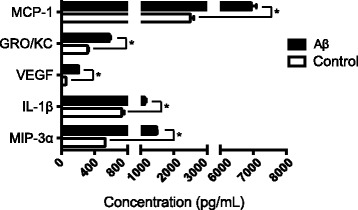
Long-Evans rats received three intravitreal injections of amyloid beta (Aβ), once every 4 days, and were sacrificed at day 14. The vitreous samples were collected to assess the levels of secreted cytokines. The inflammasome activity was evaluated by both immunohistochemistry and western blot. The types of RPE cell death mechanisms were determined using specific cell death markers and morphological characterizations.
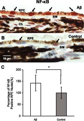
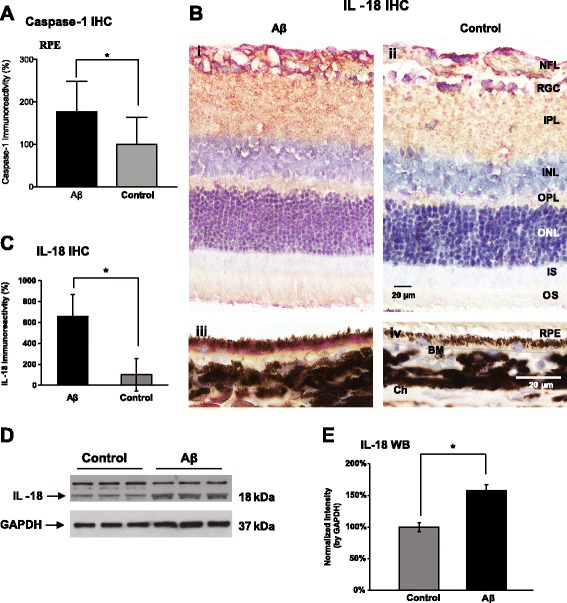
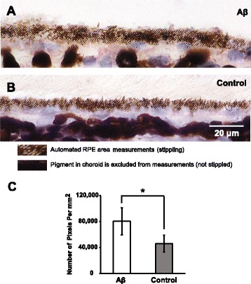
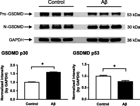
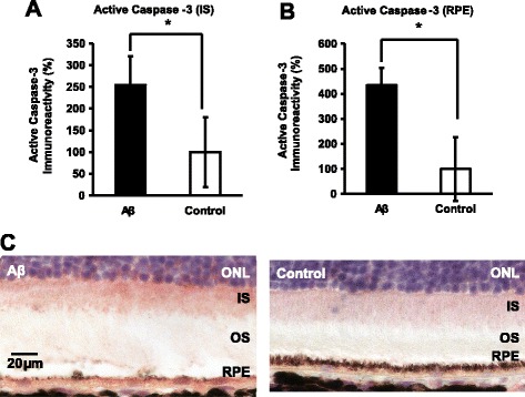
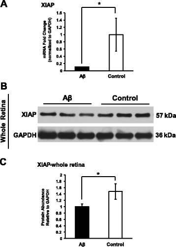
We found robust inflammasome activation evident by enhanced caspase-1 immunoreactivity, augmented NF-κB nuclear translocalization, increased IL-1β vitreal secretion, and IL-18 protein levels. Moreover, we observed elevated proteolytic cleavage of caspase-3 and gasdermin D, markers for apoptosis and pyroptosis, respectively, in RPE-choroid tissues. There was also a significant reduction in the anti-apoptotic factor, X-linked inhibitor of apoptosis protein, consistent with the overall changes of RPE cells. Morphological analysis showed phenotypic characteristics of pyroptosis including RPE cell swelling.
Our data suggest that two cell death pathways, pyroptosis and apoptosis, were activated in RPE cells after exposure to prolonged inflammasome activation, induced by a drusen component, Aβ. The involvement of two distinct cell death pathways in RPE sheds light on the potential interplay between these pathways and provides insights on the future development of therapeutic strategies for AMD.
年龄相关性黄斑变性(AMD)是一种破坏性眼病,可导致老年人视力不可逆转地丧失。视网膜色素上皮(RPE)是 AMD 中受影响的主要细胞类型,在疾病的晚期会发生程序性细胞死亡。然而,AMD 中 RPE 变性的确切机制仍未解决。目前的理论认为,每种细胞死亡途径都是独立作用的,彼此之间没有调节作用。在我们之前的研究中,我们在体内诱导了短暂的炎症小体活性爆发,现在我们研究了延长炎症小体活性对大鼠 RPE 细胞死亡机制的影响。
长耳大鼠接受三次玻璃体内注射淀粉样β(Aβ),每 4 天一次,并在第 14 天处死。收集玻璃体样本以评估分泌细胞因子的水平。通过免疫组织化学和 Western blot 评估炎症小体的活性。通过特定的细胞死亡标志物和形态特征来确定 RPE 细胞死亡机制的类型。
我们发现炎症小体激活明显,表现为 caspase-1 免疫反应性增强、NF-κB 核易位增加、IL-1β 玻璃体分泌增加和 IL-18 蛋白水平升高。此外,我们观察到 RPE-脉络膜组织中 caspase-3 和 gasdermin D 的蛋白水解切割增加,分别为凋亡和焦亡的标志物,并且 X 连锁凋亡抑制蛋白(一种抗凋亡因子)的水平显著降低,与 RPE 细胞的整体变化一致。形态学分析显示出焦亡的表型特征,包括 RPE 细胞肿胀。
我们的数据表明,两种细胞死亡途径,即焦亡和凋亡,在 RPE 细胞暴露于持续性炎症小体激活后被激活,这是由 Aβ 等 drusen 成分诱导的。两种不同的细胞死亡途径在 RPE 中的参与揭示了这些途径之间的潜在相互作用,并为 AMD 治疗策略的未来发展提供了新的见解。