Shaban Baratollah, Khajavi Amin, Khaki Nasim, Mohiti Yones, Mehri Tahere, Kermani Hamed
Department of Oral and Maxillofacial Surgery, Faculty of Dentistry, Mashhad University of Medical Sciences, Mashhad, Iran.
Department of Periodontics, Faculty of Dentistry, Mashhad University of Medical Sciences, Mashhad, Iran.
J Korean Assoc Oral Maxillofac Surg. 2017 Dec;43(6):395-400. doi: 10.5125/jkaoms.2017.43.6.395. Epub 2017 Dec 26.
The aim of this study was to evaluate different anatomical variants of the anterior loop of the inferior alveolar nerve (IAN) via cone-beam computed tomography (CBCT).
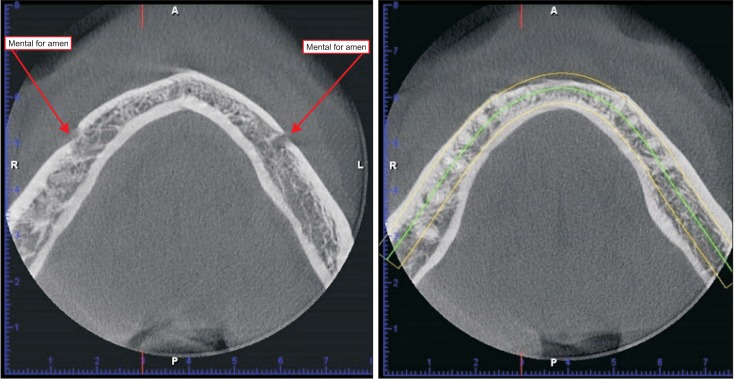
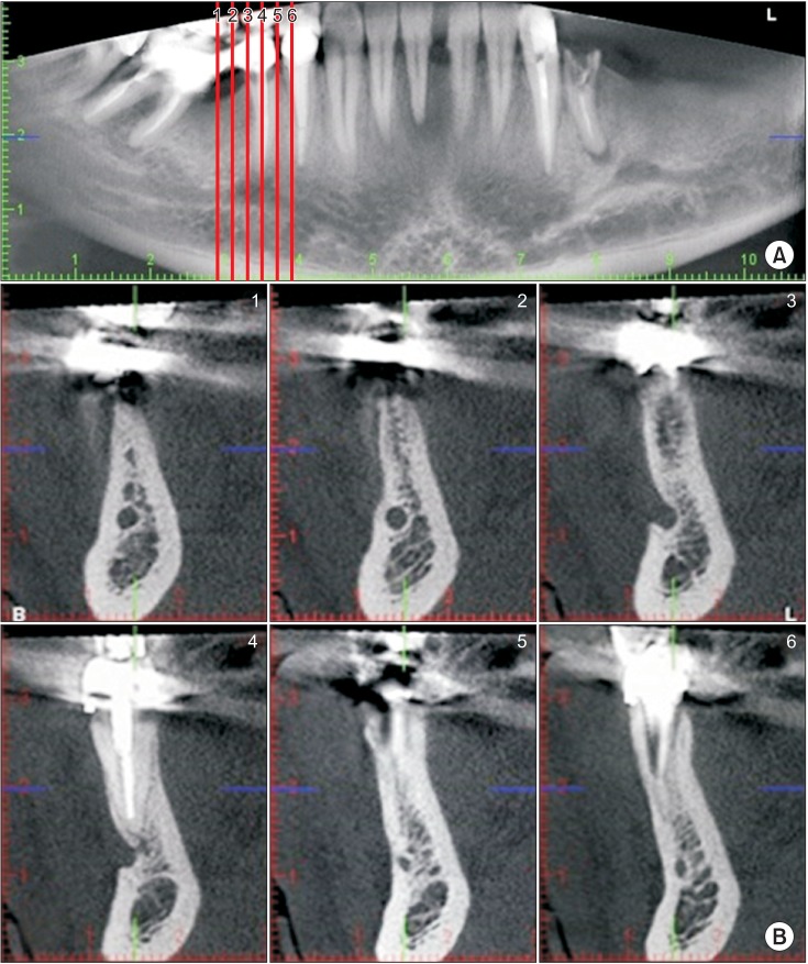
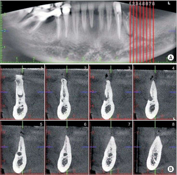
CBCT images of 71 patients (36 males and 35 females) were evaluated. We used the classification described by Solar for IAN evaluation. In this classification, three different types of IAN loops were introduced prior to emerging from the mental foramen. We classified patients according to this system and introduced a new, fourth type.
Type I was seen in 15 sites (10.6%), type II in 39 sites (27.5%), and type III in 50 sites (35.2%). We found a new type in 38 sites (26.8%) that constituted a fourth type.
We found that type III was the most common variant. In the fourth type, the IAN was not detectable because the main nerve was adjacent to the cortical plate and the incisive branch was thinner than the main branch and alongside it. In this type, more care is needed for surgeries including inferior alveolar and mental nerve transposition.
本研究旨在通过锥形束计算机断层扫描(CBCT)评估下牙槽神经(IAN)前袢的不同解剖变异。
对71例患者(36例男性和35例女性)的CBCT图像进行评估。我们采用Solar描述的分类方法进行IAN评估。在该分类中,IAN在穿出颏孔之前有三种不同类型的袢。我们根据该系统对患者进行分类,并引入了一种新的第四类型。
I型在15个部位(10.6%)出现,II型在39个部位(27.5%)出现,III型在50个部位(35.2%)出现。我们在38个部位(26.8%)发现了一种构成第四类型的新类型。
我们发现III型是最常见的变异类型。在第四类型中,由于主神经与皮质板相邻且切牙支比主支细并与之并行,IAN无法检测到。在这种类型中,包括下牙槽神经和颏神经移位在内的手术需要更加小心。