Thompson Cayla A, DeLaForest Ann, Battle Michele A
Department of Cell Biology, Neurobiology, and Anatomy, Medical College of Wisconsin, 8701 Watertown Plank Road, Milwaukee, WI 53226, USA.
Department of Cell Biology, Neurobiology, and Anatomy, Medical College of Wisconsin, 8701 Watertown Plank Road, Milwaukee, WI 53226, USA.
Dev Biol. 2018 Mar 15;435(2):97-108. doi: 10.1016/j.ydbio.2018.01.006. Epub 2018 Jan 12.
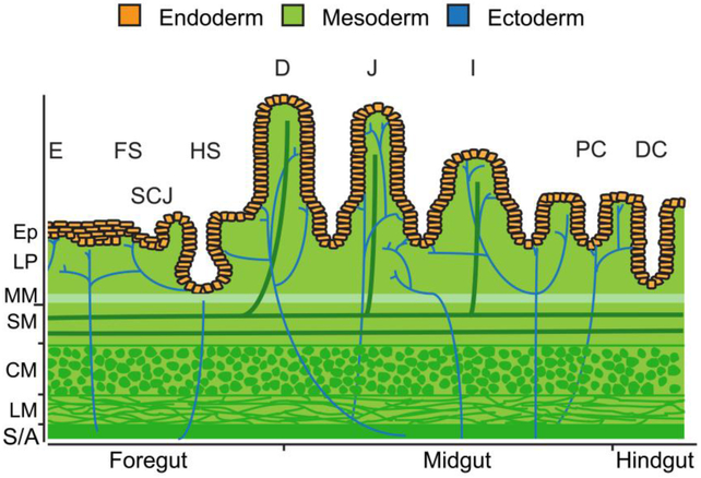
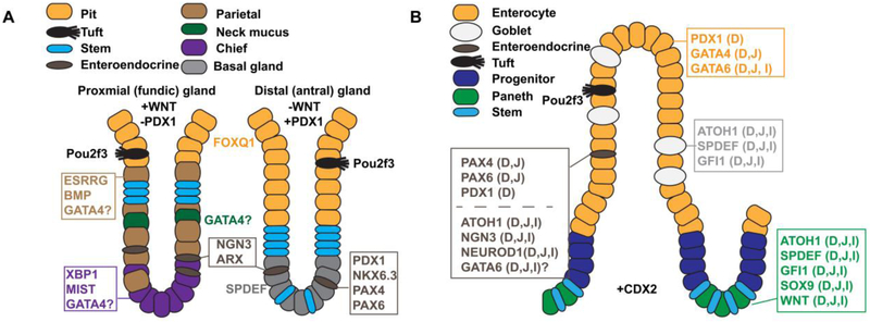
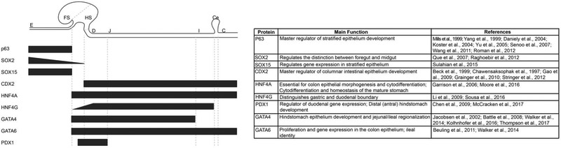
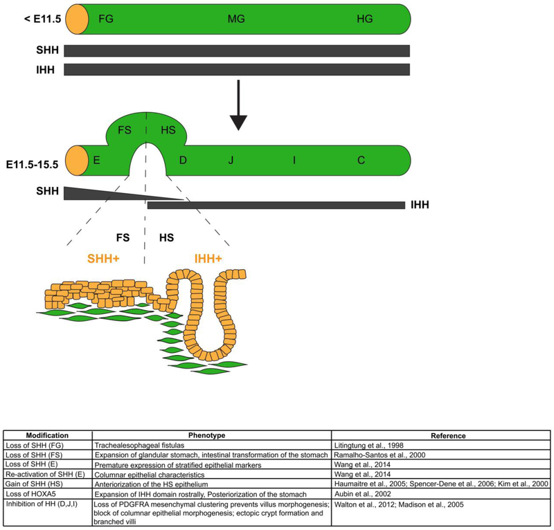
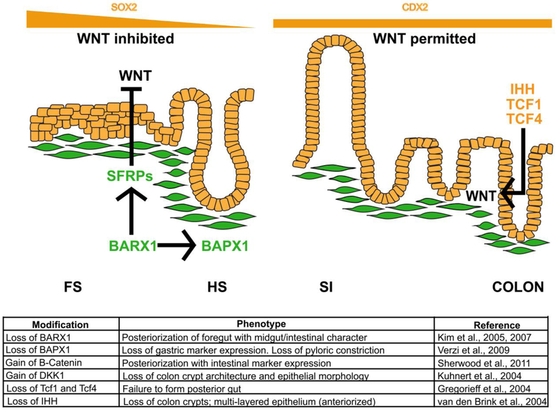
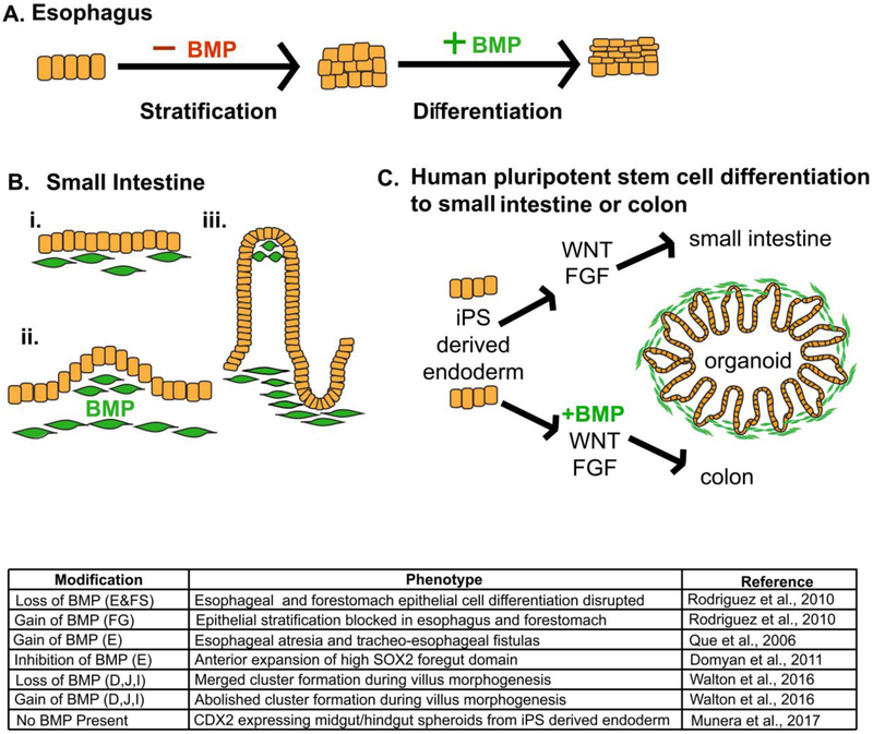
The gastrointestinal (GI) tract, in simplest terms, can be described as an epithelial-lined muscular tube extending along the cephalocaudal axis from the oral cavity to the anus. Although the general architecture of the GI tract organs is conserved from end to end, the presence of different epithelial tissue structures and unique epithelial cell types within each organ enables each to perform the distinct digestive functions required for efficient nutrient assimilation. Spatiotemporal regulation of signaling pathways and downstream transcription factors controls GI epithelial morphogenesis during development to confer essential regional-specific epithelial structures and functions. Here, we discuss the fundamental functions of each GI tract organ and summarize the diversity of epithelial structures present along the cephalocaudal axis of the GI tract. Next, we discuss findings, primarily from genetic mouse models, that have defined the roles of key transcription factors during epithelial morphogenesis, including p63, SOX2, SOX15, GATA4, GATA6, HNF4A, and HNF4G. Additionally, we examine how the Hedgehog, WNT, and BMP signaling pathways contribute to defining unique epithelial features along the cephalocaudal axis of the GI tract. Lastly, we examine the molecular mechanisms controlling regionalized cytodifferentiation of organ-specific epithelial cell types within the GI tract, concentrating on the stomach and small intestine. The delineation of GI epithelial patterning mechanisms in mice has provided fundamental knowledge to guide the development and refinement of three-dimensional GI organotypic culture models such as those derived from directed differentiation of human pluripotent stem cells and those derived directly from human tissue samples. Continued examination of these pathways will undoubtedly provide vital insights into the mechanisms of GI development and disease and may afford new avenues for innovative tissue engineering and personalized medicine approaches to treating GI diseases.
简而言之,胃肠道(GI)可被描述为一条内衬上皮的肌性管道,沿头尾轴从口腔延伸至肛门。尽管胃肠道器官的总体结构从头到尾是保守的,但每个器官内不同的上皮组织结构和独特的上皮细胞类型使其能够执行有效营养吸收所需的独特消化功能。信号通路和下游转录因子的时空调节在发育过程中控制胃肠道上皮形态发生,以赋予基本的区域特异性上皮结构和功能。在这里,我们讨论每个胃肠道器官的基本功能,并总结沿胃肠道头尾轴存在的上皮结构的多样性。接下来,我们讨论主要来自基因小鼠模型的研究结果,这些结果确定了关键转录因子在上皮形态发生过程中的作用,包括p63、SOX2、SOX15、GATA4、GATA6、HNF4A和HNF4G。此外,我们研究刺猬信号通路、WNT信号通路和骨形态发生蛋白(BMP)信号通路如何有助于定义沿胃肠道头尾轴的独特上皮特征。最后,我们研究控制胃肠道内器官特异性上皮细胞类型区域化细胞分化的分子机制,重点关注胃和小肠。小鼠胃肠道上皮模式形成机制的描述为指导三维胃肠道器官型培养模型的开发和完善提供了基础知识,例如那些源自人多能干细胞定向分化的模型以及直接源自人体组织样本的模型。对这些信号通路的持续研究无疑将为胃肠道发育和疾病的机制提供重要见解,并可能为治疗胃肠道疾病的创新组织工程和个性化医疗方法提供新途径。