Division of Gynecologic Oncology, Department of Obstetrics and Gynecology, Comprehensive Cancer Center, the Ohio State University Wexner Medical Center, Columbus, Ohio.
Department of Pathology, Gynecological Pathology and Cytopathology Unit, the Ohio State University Wexner Medical Center, Columbus, Ohio.
Cancer Res. 2018 Apr 1;78(7):1739-1750. doi: 10.1158/0008-5472.CAN-17-1671. Epub 2018 Jan 16.
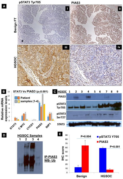
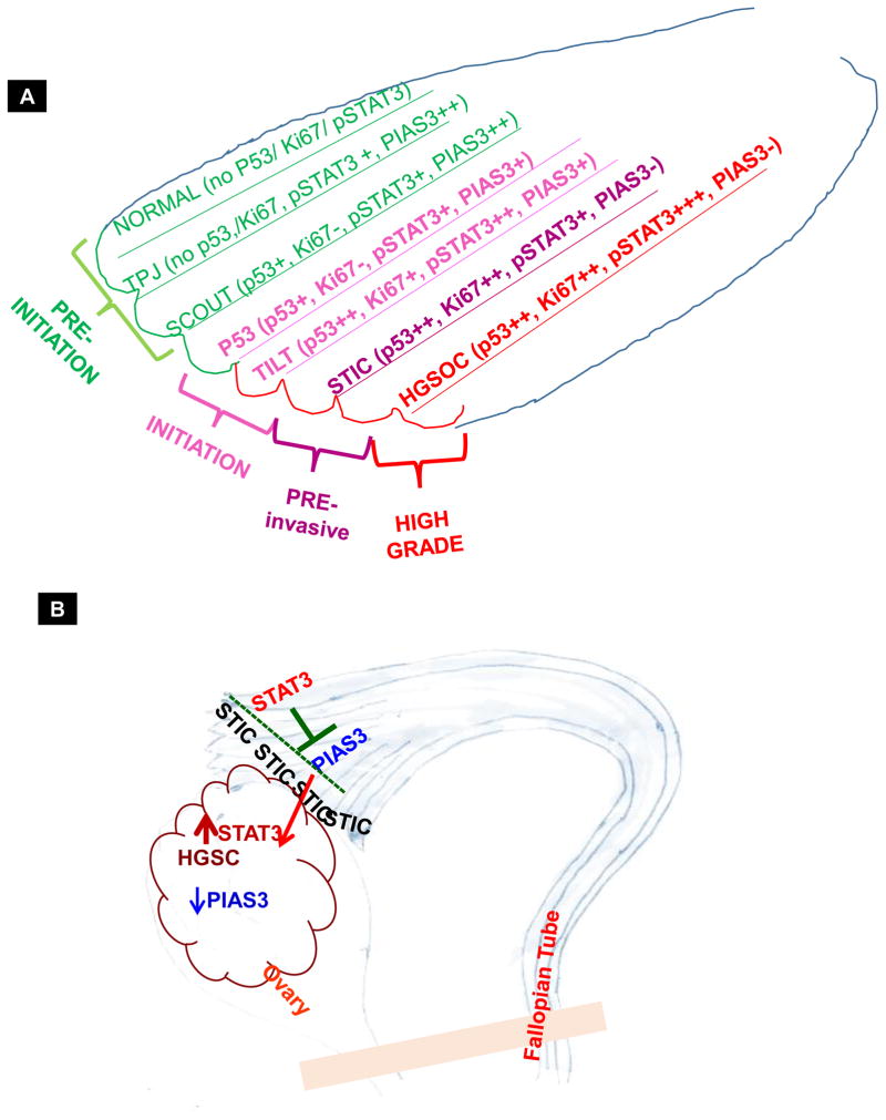
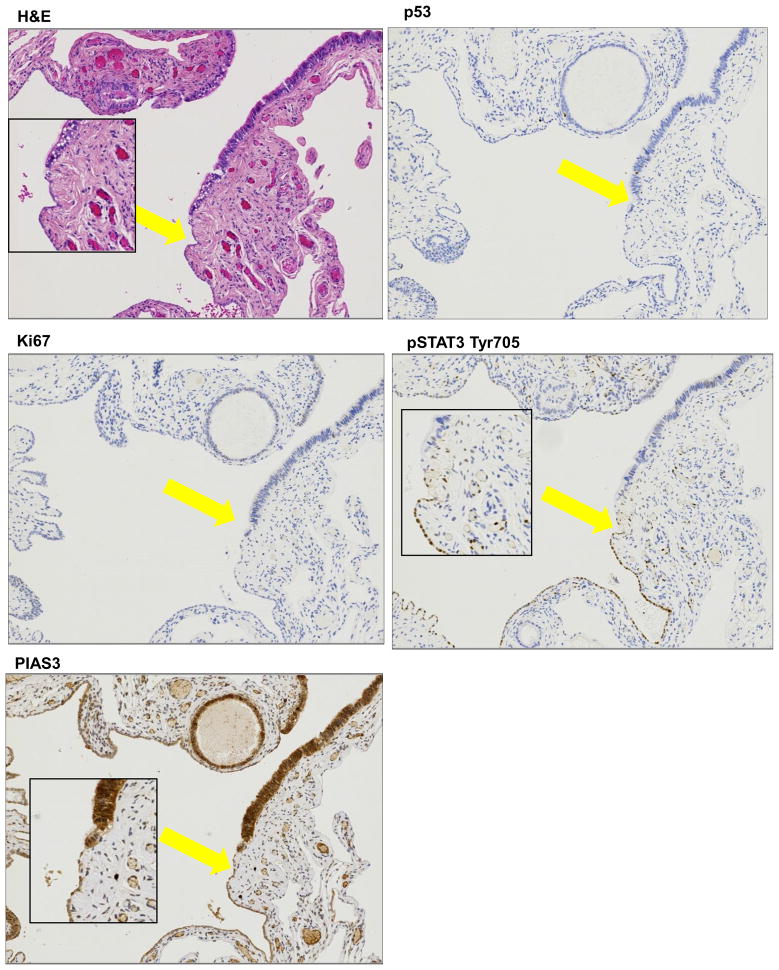
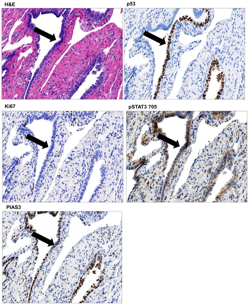
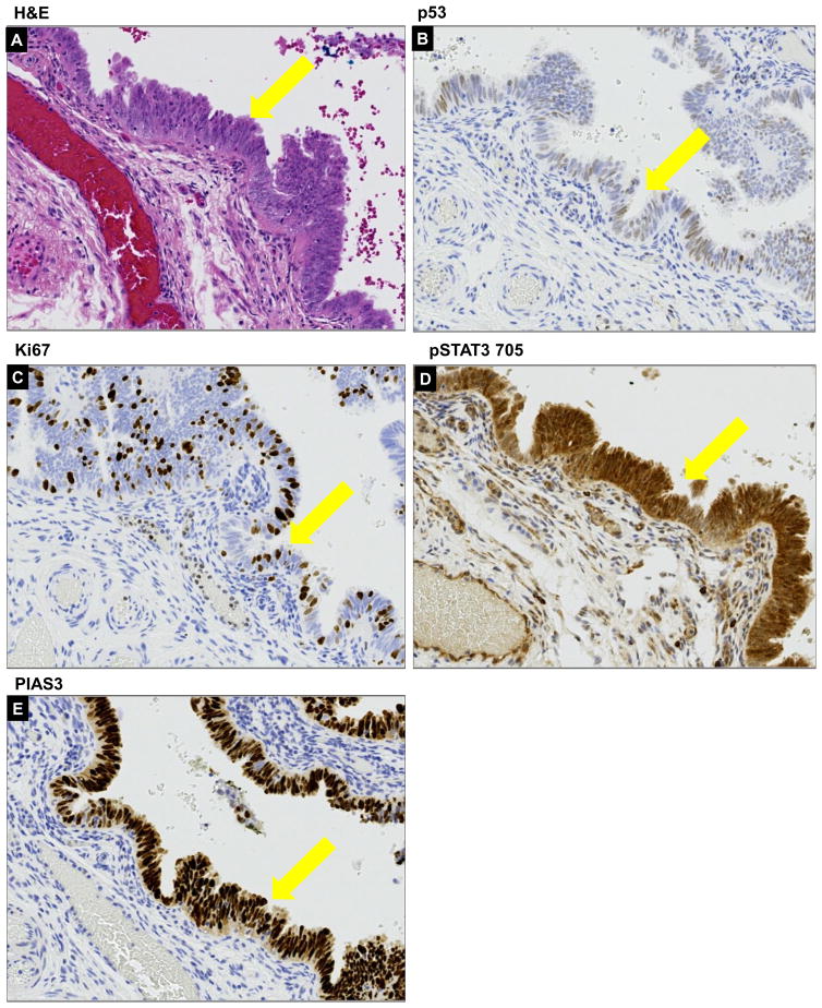
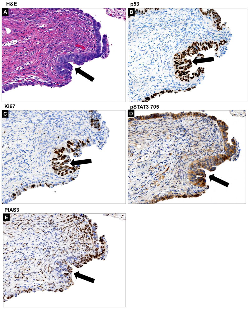
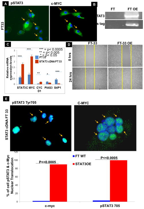
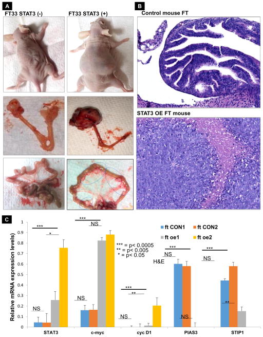
The initial molecular events that lead to malignant transformation of the fimbria of the fallopian tube (FT) through high-grade serous ovarian carcinoma (HGSC) remain poorly understood. In this study, we report that increased expression of signal transducer and activator of transcription 3 () and suppression or loss of protein inhibitor of activated STAT3 () in FT likely drive HGSC. We evaluated human tissues-benign normal FT, tubal-peritoneal junction (TPJ), p53 signature FT tissue, tubal intraepithelial lesion in transition (TILT), serous tubal intraepithelial carcinoma (STIC) without ovarian cancer, and HGSC for expression of (compared with their known signature) and their target proliferation genes. We observed constitutive activation of and low levels or loss of in the TPJ, p53 signature, TILT, and STIC through advanced stage IV (HGSC) tissues. Elevated expression of and decreased levels of appeared as early as TPJ and the trend continued until very advanced stage HGSC (compared with high and low expression in normal benign FT). Exogenous expression of in FT cells mediated translocation of and into the nucleus. experiments demonstrated that overexpression of in FT secretory epithelial cells promoted tumor progression and metastasis, mimicking the clinical disease observed in patients with HGSC. Thus, we conclude that the pathway plays a role in the development and progression of HGSC from its earliest premalignant states. Concomitant gain of pSTAT3 Tyr705 and loss of PIAS3 appear critical for initiation and development of high-grade serous carcinoma. .
导致输卵管(FT)纤毛恶性转化的最初分子事件,通过高级别浆液性卵巢癌(HGSC)仍然知之甚少。在这项研究中,我们报告说,信号转导和转录激活因子 3(STAT3)的表达增加和蛋白抑制剂激活 STAT3(PIAS3)的抑制或缺失,可能导致 HGSC。我们评估了人组织-良性正常 FT、输卵管-腹膜交界(TPJ)、p53 特征性 FT 组织、过渡性输卵管上皮内病变(TILT)、无卵巢癌的浆液性输卵管上皮内癌(STIC)和 HGSC 中表达的(与已知的 STAT3 特征相比)及其靶增殖基因。我们观察到 TPJ、p53 特征、TILT 和 STIC 中的 STAT3 持续激活和低水平或缺失,直至晚期 IV 期(HGSC)组织。在 TPJ 和趋势一直持续到非常晚期的 HGSC(与正常良性 FT 中的高 表达和低 表达相比)中,出现了 表达升高和 水平降低。FT 细胞中 的外源性表达介导了 易位到细胞核。实验表明,FT 分泌上皮细胞中 的过表达促进了肿瘤的进展和转移,模拟了在 HGSC 患者中观察到的临床疾病。因此,我们得出结论,STAT3 途径在 HGSC 从最早的癌前状态发展和进展中起作用。同时获得 pSTAT3 Tyr705 和失去 PIAS3 似乎对高级别浆液性癌的发生和发展至关重要。