Xia Pan Pan, Li Lian Jing, Qi Run Di, Shi Jiao Jiao, Ju Wei Zhu, Chen Ming Long
Department of Cardiology, The First Affiliated Hospital of Nanjing Medical University; Nanjing-China.
Anatol J Cardiol. 2018 Mar;19(3):169-175. doi: 10.14744/AnatolJCardiol.2017.7844. Epub 2018 Jan 17.
Hypertension is a significant risk factor for atrial fibrillation (AF). The role of pulmonary vein (PV) remodeling in the mechanistic association between hypertension and AF is not definitive. In this study, we aimed to identify changes in the electrophysiology and histology in PVs in two-kidney, one-clip (2K1C) hypertensive rats.
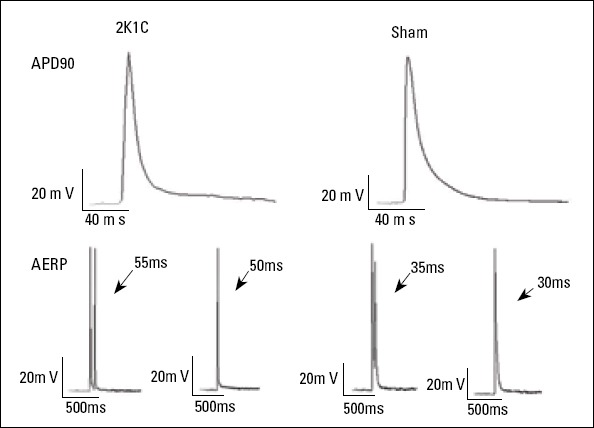
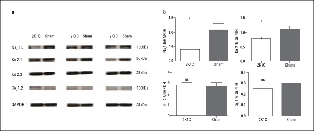
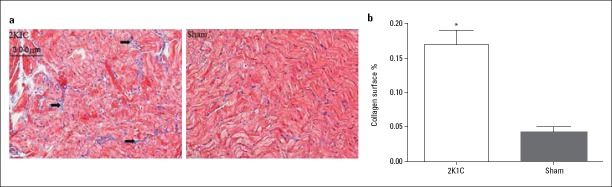
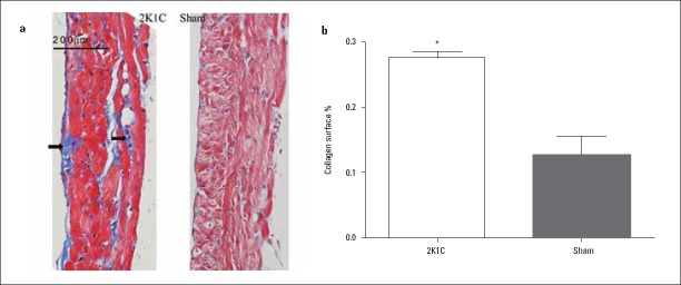
Fifty male Sprague-Dawley rats were classified into the 2K1C and sham-operated groups. The systolic blood pressure was measured every 2 weeks. The left atrial diameter was measured by transthoracic echocardiography. Left superior PV (LSPV) and left atrial (LA) fibrosis was evaluated by Masson's trichrome staining. The expression of fibrosis markers [angiotensin II (Ang II), transforming growth factor-ß1 (TGF-ß1), matrix metalloproteinase-2 (MMP-2), and collagen I (Col I)] and ion channels [Kir2.1, Kir2.3, Cav1.2, and Nav1.5] in LSVP was quantified by western blot. Conventional microelectrodes were used to record the action potential duration at 90% repolarization (APD90) and effective refractory period (ERP) in isolated LA.
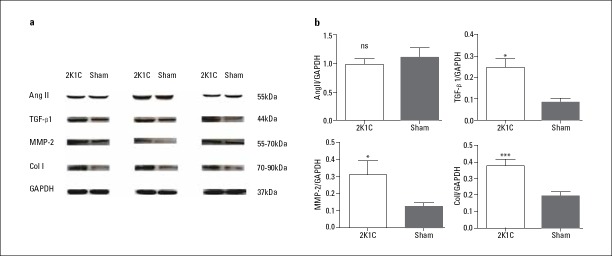
At 4 months, the 2K1C hypertensive rats developed LA dilation. Col deposition in LSPV and left atrium and expression of TGF-ß1, MMP-2, and Col I in LSPV were significantly increased in 2K1C hypertensive rats. In addition, hypertension reduced the expression of Nav1.5 and Kir2.1, although there were no significant differences in APD90; ERP; and expression of Ang II, Kir2.3, and Cav1.2 between the two groups.
Hypertension may lead to changes in the electrophysiology and histology of rats PVs, which is characterized by significant reduction in the expression of Nav1.5 and Kir2.1 and increase in interstitial fibrosis. These observations may clarify the role of PVs in the mechanistic association between hypertension and AF.
高血压是心房颤动(AF)的一个重要危险因素。肺静脉(PV)重塑在高血压与AF机制关联中的作用尚不明确。在本研究中,我们旨在确定二肾一夹(2K1C)高血压大鼠肺静脉的电生理学和组织学变化。
将50只雄性Sprague-Dawley大鼠分为2K1C组和假手术组。每2周测量收缩压。经胸超声心动图测量左心房直径。采用Masson三色染色评估左上肺静脉(LSPV)和左心房(LA)纤维化。通过蛋白质印迹法定量分析LSPV中纤维化标志物[血管紧张素II(Ang II)、转化生长因子-β1(TGF-β1)、基质金属蛋白酶-2(MMP-2)和I型胶原(Col I)]和离子通道[Kir2.1、Kir2.3、Cav1.2和Nav1.5]的表达。使用传统微电极记录离体左心房90%复极化时的动作电位时程(APD90)和有效不应期(ERP)。
4个月时,2K1C高血压大鼠出现左心房扩张。2K1C高血压大鼠LSPV和左心房中的胶原沉积以及LSPV中TGF-β1、MMP-2和Col I的表达显著增加。此外,高血压降低了Nav1.5和Kir2.1的表达,尽管两组之间的APD90、ERP以及Ang II、Kir2.3和Cav1.2的表达没有显著差异。
高血压可能导致大鼠肺静脉电生理学和组织学变化,其特征是Nav1.5和Kir2.1表达显著降低以及间质纤维化增加。这些观察结果可能阐明肺静脉在高血压与AF机制关联中的作用。