Division of Neuroscience and Experimental Psychology, School of Biological Sciences, Faculty of Biology, Medicine and Health, University of Manchester, Manchester, UK.
Stem Cell Res Ther. 2018 Jan 17;9(1):11. doi: 10.1186/s13287-017-0753-5.
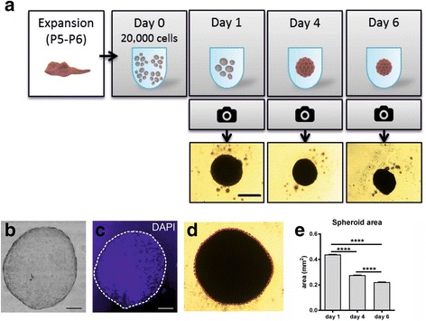
Mesenchymal stem cells (MSCs) are one of the most promising candidates for the treatment of major neurological disorders. Desirable therapeutic properties of MSCs include reparative and regenerative potential but, despite their proven safety, the efficacy of MSCs remains controversial. Therefore, it is essential to optimise culture protocols to enhance the therapeutic potential of the MSC secretome. Here we aimed to: assess the increase in secretion of cytokines that may induce repair, regeneration, or immunomodulation when cultured in three dimensions; study the effect of interleukin (IL)-1 priming on two- (2D) and three-dimensional (3D) cultures of MSC; and evaluate the potential use of the modified secretome using microglial-MSC co-cultures.
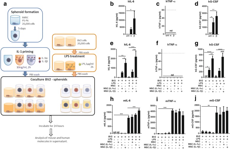
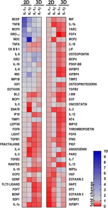
We established a 3D spheroid culture of human MSCs, and compared the secretome in 2D and 3D cultures under primed (IL-1) and unprimed conditions. BV2 microglial cells were stimulated with lipopolysaccharide (LPS) and treated with spheroid conditioned media (CM) or were co-cultured with whole spheroids. Concentrations of secreted cytokines were determined by enzyme-linked immunosorbent assay (ELISA). Protein arrays were used to further evaluate the effect of IL-1 priming in 2D and 3D cultures.
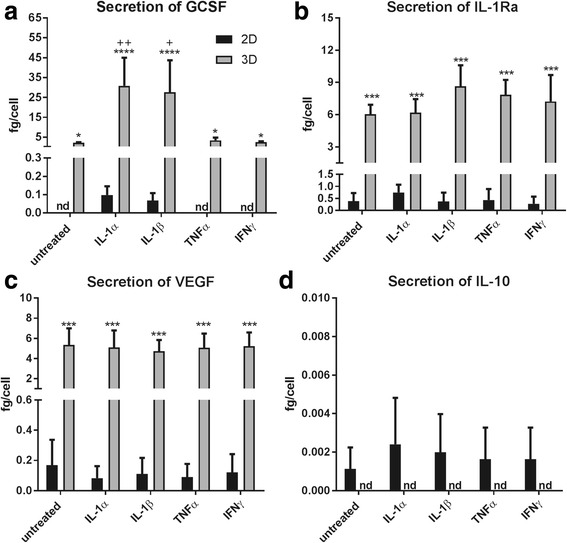
3D culture of MSCs significantly increased secretion of the IL-1 receptor antagonist (IL-1Ra), vascular endothelial growth factor (VEGF), and granulocyte-colony stimulating factor (G-CSF) compared with 2D culture, despite priming treatments with IL-1 being more effective in 2D than in 3D. The addition of CM of 3D-MSCs reduced LPS-induced tumour necrosis factor (TNF)-α secretion from BV2 cells, while the 3D spheroid co-cultured with the BV2 cells induced an increase in IL-6, but had no effect on TNF-α release. Protein arrays indicated that priming treatments trigger a more potent immune profile which is necessary to orchestrate an effective tissue repair. This effect was lost in 3D, partly because of the overexpression of IL-6.
Increased secretion of anti-inflammatory markers occurs when MSCs are cultured in 3D, but this specific secretome did not translate into anti-inflammatory effects on LPS-treated BV2 cells in co-culture. These data highlight the importance of optimising priming treatments and culture conditions to maximise the therapeutic potential of MSC spheroids.
间充质干细胞(MSCs)是治疗重大神经疾病最有希望的候选者之一。MSCs 的理想治疗特性包括修复和再生潜能,但尽管已证明其安全性,但其疗效仍存在争议。因此,优化培养方案以增强 MSC 分泌组的治疗潜力至关重要。在这里,我们旨在:评估在三维培养时分泌细胞因子的增加,这些细胞因子可能诱导修复、再生或免疫调节;研究白细胞介素(IL)-1 引发对 MSC 二维(2D)和三维(3D)培养的影响;并使用微胶质细胞-MSC 共培养来评估改良分泌组的潜在用途。
我们建立了人 MSC 的 3D 球体培养,并比较了在有(IL-1)和无引发条件下 2D 和 3D 培养中的分泌组。用脂多糖(LPS)刺激 BV2 小胶质细胞,并用球体条件培养基(CM)处理或与整个球体共培养。通过酶联免疫吸附测定(ELISA)测定分泌细胞因子的浓度。蛋白质阵列用于进一步评估 IL-1 引发在 2D 和 3D 培养中的作用。
与 2D 培养相比,MSC 的 3D 培养显著增加了白细胞介素 1 受体拮抗剂(IL-1Ra)、血管内皮生长因子(VEGF)和粒细胞集落刺激因子(G-CSF)的分泌,尽管在 2D 中用 IL-1 引发比在 3D 中更有效。3D-MSC 的 CM 添加减少了 LPS 诱导的 BV2 细胞肿瘤坏死因子(TNF)-α的分泌,而与 BV2 细胞共培养的 3D 球体诱导了 IL-6 的增加,但对 TNF-α的释放没有影响。蛋白质阵列表明,引发处理引发了更有效的免疫谱,这对于协调有效的组织修复是必要的。在 3D 中,这种效应丢失了,部分原因是 IL-6 的过度表达。
当 MSCs 在 3D 中培养时,抗炎标志物的分泌增加,但这种特定的分泌组在共培养的 LPS 处理的 BV2 细胞中没有转化为抗炎作用。这些数据强调了优化引发处理和培养条件以最大化 MSC 球体治疗潜力的重要性。