Leduc Charlotte, Adam Julien, Louvet Emilie, Sourisseau Tony, Dorvault Nicolas, Bernard Marine, Maingot Elodie, Faivre Laura, Cassin-Kuo Mei-Shiue, Boissier Emilie, Dessoliers Marie-Charlotte, Robin Angélique, Casiraghi Odile, Even Caroline, Temam Stéphane, Olaussen Ken A, Soria Jean-Charles, Postel-Vinay Sophie
INSERM, UMR981, F-94805, Villejuif, France, Paris Sud University, Gustave Roussy, Villejuif, France.
Department of Pathology, Gustave Roussy, Villejuif, France.
ESMO Open. 2018 Jan 9;3(1):e000257. doi: 10.1136/esmoopen-2017-000257. eCollection 2018.
Antiprogrammed cell death-1/programmed cell death-ligand 1 (PD-1/PD-L1) therapies have demonstrated promising activity in advanced head and neck squamous cell carcinoma (HNSCC), with overall response rates of approximately 20% in unselected populations and survival benefit. Whether induction docetaxel, platinum and fluorouracil (TPF) modifies PD-L1 expression or tumour immune infiltrates is unknown.
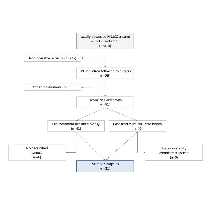
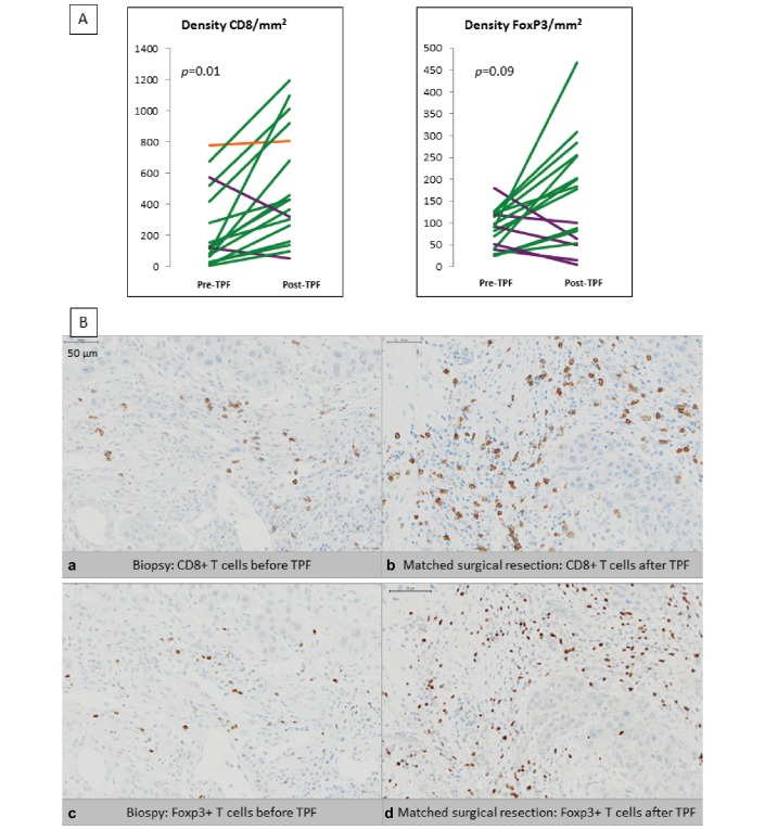
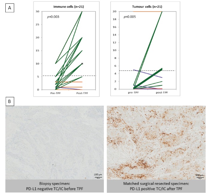
Patients with locally advanced HNSCC treated at Gustave Roussy (Villejuif, France) between 2006 and 2013 by induction TPF followed by surgery were retrospectively considered. Patients with paired samples (pre-TPF and post-TPF) were kept for further analysis. PD-L1 expression was quantified by immunohistochemistry according to a validated protocol. The objective of the study was to compare PD-L1 expression on tumour cells (TC) and immune cells (IC) (positivity threshold of ≥5%) before and after TPF. CD8+ and Foxp3+ lymphocytes densities before and after TPF were also quantified.
Out of 313 patients receiving induction TPF, 86 underwent surgery; paired samples were available for 21 of them. Baseline PD-L1 expression was ≥5% in two and five samples for TC and IC, respectively. A significant increase of PD-L1 expression was observed after TPF, with 15 samples (71%) presenting a positive staining in IC after induction chemotherapy (P=0.003; Wilcoxon rank-sum test) and eight samples (38%) in TC (P=0.005; Wilcoxon rank-sum test). Tumour-infiltrating CD8+ mean densities also significantly increased post-TPF (P=0.01). There was no significant difference in Foxp3+ expression, CD8/Foxp3 ratio or correlation with outcome.
TPF induction chemotherapy in advanced HNSCC increases PD-L1 positivity on tumour-infiltrating ICs, as well as CD8+ lymphocytes density. These results warrant independent validation on larger datasets and might help therapeutic strategy in advanced HNSCC.
抗程序性细胞死亡蛋白1/程序性细胞死亡配体1(PD-1/PD-L1)疗法在晚期头颈部鳞状细胞癌(HNSCC)中已显示出有前景的活性,在未选择的人群中总体缓解率约为20%,且有生存获益。诱导多西他赛、铂类和氟尿嘧啶(TPF)是否会改变PD-L1表达或肿瘤免疫浸润尚不清楚。
回顾性分析2006年至2013年在法国维勒瑞夫古斯塔夫·鲁西研究所接受诱导TPF治疗后行手术的局部晚期HNSCC患者。保留有配对样本(TPF治疗前和治疗后)的患者进行进一步分析。根据经过验证的方案通过免疫组织化学对PD-L1表达进行定量。本研究的目的是比较TPF治疗前后肿瘤细胞(TC)和免疫细胞(IC)上的PD-L1表达(阳性阈值≥5%)。还对TPF治疗前后CD8+和Foxp3+淋巴细胞密度进行了定量。
在313例接受诱导TPF治疗的患者中,86例行手术;其中21例有配对样本。基线时,TC和IC的PD-L1表达分别在2例和5例样本中≥5%。TPF治疗后观察到PD-L1表达显著增加,诱导化疗后15例样本(71%)的IC呈阳性染色(P=0.003;Wilcoxon秩和检验),8例样本(38%)的TC呈阳性染色(P=0.005;Wilcoxon秩和检验)。TPF治疗后肿瘤浸润性CD8+平均密度也显著增加(P=0.01)。Foxp3+表达、CD8/Foxp3比值或与预后的相关性无显著差异。
晚期HNSCC的TPF诱导化疗增加了肿瘤浸润ICs上的PD-L1阳性率以及CD8+淋巴细胞密度。这些结果需要在更大的数据集中进行独立验证,可能有助于晚期HNSCC的治疗策略制定。