Department of Orthopedic Surgery, Yijishan Hospital, The First Affiliated Hospital of Wannan Medical College, Wuhu, Anhui 241001, P.R. China.
Department of Surgery, Yijishan Hospital, The First Affiliated Hospital of Wannan Medical College, Wuhu, Anhui 241001, P.R. China.
Mol Med Rep. 2018 Mar;17(3):4415-4421. doi: 10.3892/mmr.2018.8435. Epub 2018 Jan 16.
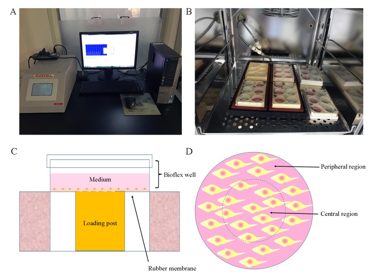
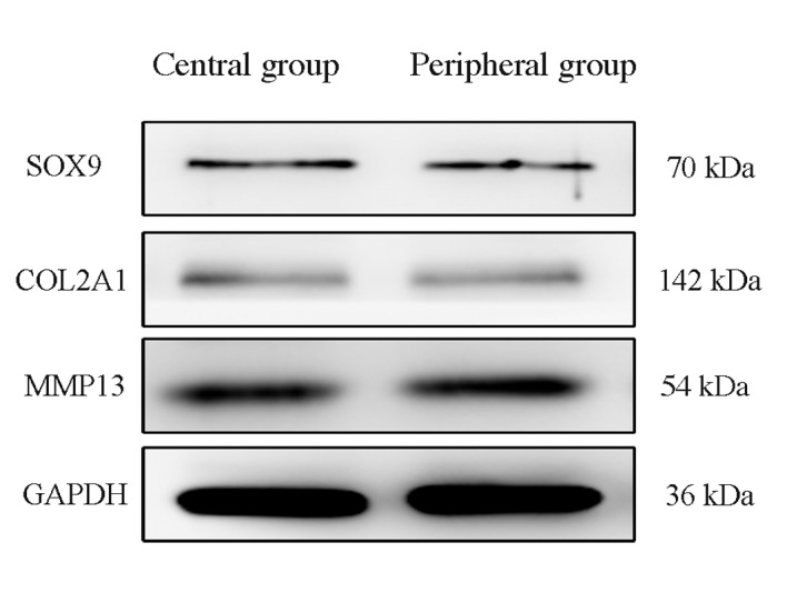
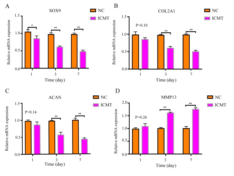
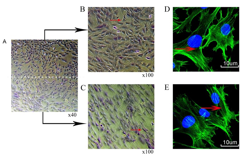
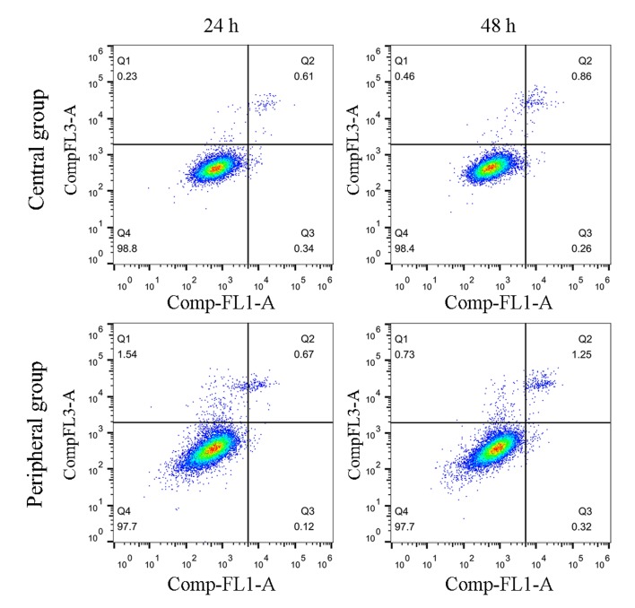
The aim of this study was to explore the degree of degeneration of endplate chondrocytes in different tension regions induced by intermittent cyclic mechanical tension (ICMT) in vitro. Rat endplate chondrocytes were harvested and treated with 10% ICMT for 8 h/day with a frequency of 0.5 Hz. A cartilage degeneration model was induced using an FX‑5000T cell strain‑loading system. The experiment was divided into the central region and the peripheral region, according to the contact area between the loading post and the six‑well flexible silicone rubber BioFlex plates. Toluidine blue and phalloidin staining were used to observe the morphological changes of cells following mechanical stimulation. Apoptosis was detected by flow cytometry and the mRNA and protein expression levels of collagen type II α1, aggrecan, SRY‑box 9 and matrix metalloproteinase 13 were detected by reverse transcription‑quantitative polymerase chain reaction (RT‑qPCR) and western blotting, respectively. Endplate chondrocytes exhibited degenerative alterations under mechanical conditions of 10% ICMT and 0.5 Hz at 8 h/day. Toluidine blue and phalloidin staining demonstrated that the cells in the peripheral region were more slender compared with cells in the central region, but RT‑qPCR and western blotting results demonstrated that the degree of cell degeneration between the two groups was not statistically differences. So that cell morphological alteration does not imply that cells have undergone degeneration.
本研究旨在探讨体外间歇循环机械张力(ICMT)在不同张应力区域引起的终板软骨细胞退变程度。从大鼠终板软骨细胞中分离并提取,以 10% ICMT 处理 8 小时/天,频率为 0.5Hz。采用 FX-5000T 细胞加载系统诱导软骨退变模型。根据加载柱与六孔柔性硅橡胶 BioFlex 板之间的接触面积,实验分为中心区域和周围区域。通过甲苯胺蓝和鬼笔环肽染色观察细胞在机械刺激后的形态变化。通过流式细胞术检测细胞凋亡,通过逆转录定量聚合酶链反应(RT-qPCR)和 Western blot 检测 II 型胶原α1、聚集蛋白聚糖、SRY 框 9 和基质金属蛋白酶 13 的 mRNA 和蛋白表达水平。终板软骨细胞在 10% ICMT 和 0.5Hz 条件下,每天 8 小时,表现出退行性改变。甲苯胺蓝和鬼笔环肽染色表明,周围区域的细胞比中心区域的细胞更细长,但 RT-qPCR 和 Western blot 结果表明,两组细胞退变程度无统计学差异。因此,细胞形态的改变并不意味着细胞已经发生退变。