Maymó Julieta L, Riedel Rodrigo, Pérez-Pérez Antonio, Magatti Marta, Maskin Bernardo, Dueñas José Luis, Parolini Ornella, Sánchez-Margalet Víctor, Varone Cecilia L
Universidad de Buenos Aires, CONICET, Instituto de Química Biológica de la Facultad de Ciencias Exactas y Naturales (IQUIBICEN), Ciudad Universitaria Pabellón 2, 4° piso, (1428), Buenos Aires, Argentina.
Universidad de Buenos Aires, Facultad de Ciencias Exactas y Naturales, Departamento de Química Biológica, Ciudad Universitaria Pabellón 2, 4° piso, (1428), Buenos Aires, Argentina.
PLoS One. 2018 Jan 18;13(1):e0191489. doi: 10.1371/journal.pone.0191489. eCollection 2018.
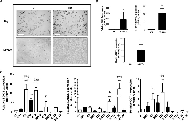
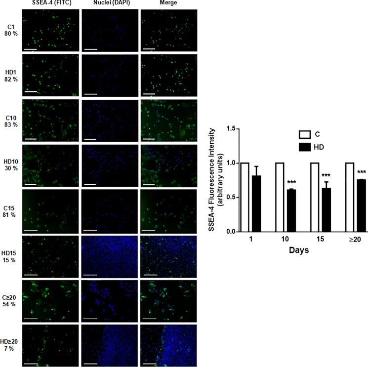
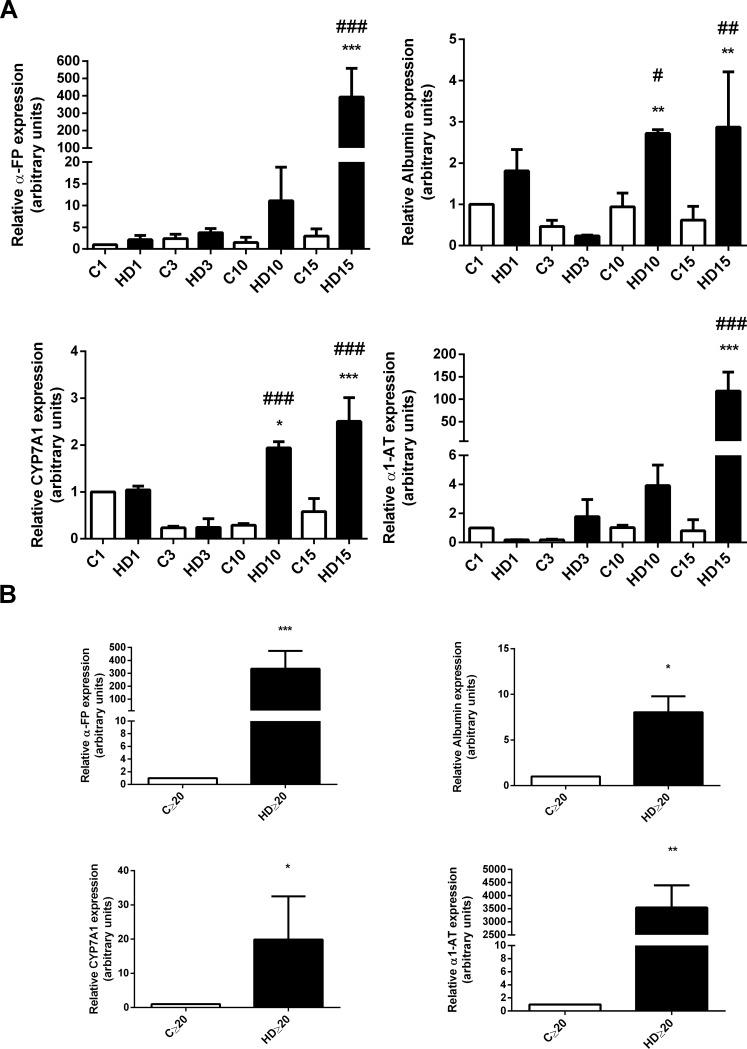
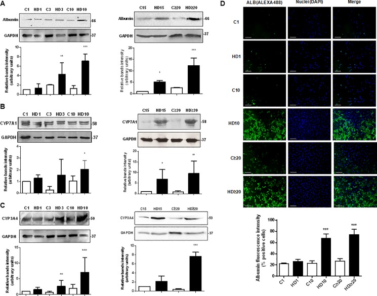
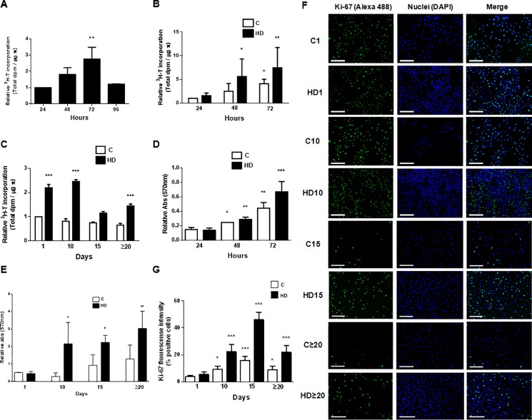
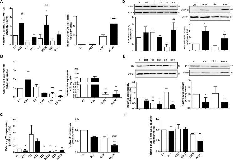
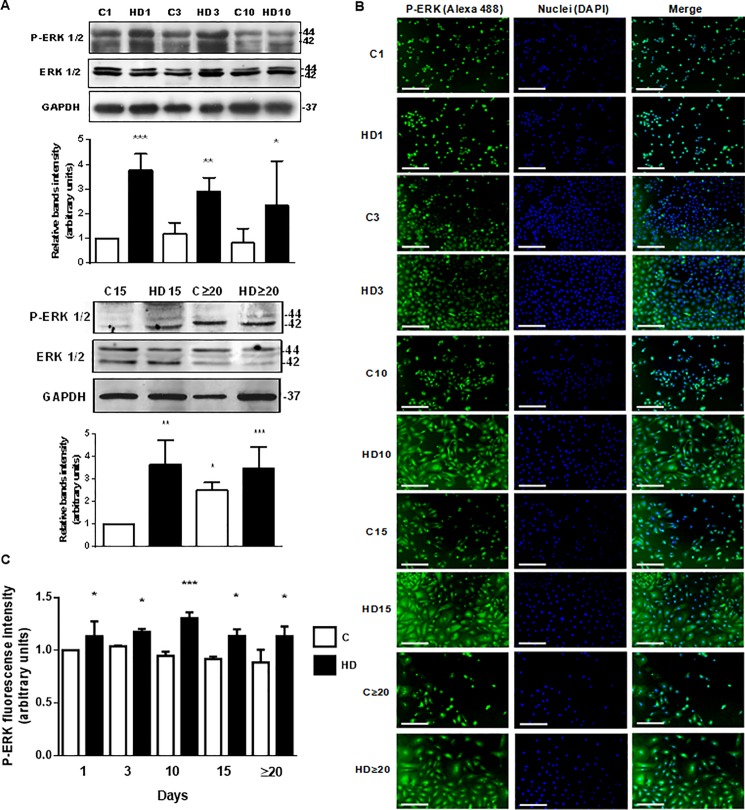
Stem cells derived from placental tissues are an attractive source of cells for regenerative medicine. Amniotic epithelial cells isolated from human amnion (hAECs) have desirable and competitive characteristics that make them stand out between other stem cells. They have the ability to differentiate toward all three germ layers, they are not tumorigenic and they have immunosuppressive properties. Although liver transplantation is the best way to treat acute and chronic hepatic failure patients, there are several obstacles. Recently, stem cells have been spotlighted as alternative source of hepatocytes because of their potential for hepatogenic differentiation. In this work, we aimed to study the proliferation and survival of the hAECs during their hepatic differentiation. We have also analyzed the changes in pluripotency and hepatic markers. We differentiated amniotic cells applying a specific hepatic differentiation (HD) protocol. We determined by qRT-PCR that hAECs express significant levels of SOX-2, OCT-4 and NANOG during at least 15 days in culture and these pluripotent markers diminish during HD. SSEA-4 expression was reduced during HD, measured by immunofluorescence. Morphological characteristics became more similar to hepatic ones in differentiated cells and representative hepatic markers significantly augmented their expression, measured by qRT-PCR and Western blot. Cells achieved a differentiation efficiency of 75%. We observed that HD induced proliferation and promoted survival of hAECs, during 30 days in culture, evaluated by 3H-thymidine incorporation and MTT assay. HD also promoted changes in hAECs cell cycle. Cyclin D1 expression increased, while p21 and p53 levels were reduced. Immunofluorescence analysis showed that Ki-67 expression was upregulated during HD. Finally, ERK 1/2 phosphorylation, which is intimately linked to proliferation and cell survival, augmented during all HD process and the inhibition of this signaling pathway affected not only proliferation but also differentiation. Our results suggest that HD promotes proliferation and survival of hAECs, providing important evidence about the mechanisms governing their hepatic differentiation. We bring new knowledge concerning some of the optimal transplantation conditions for these hepatic like cells.
源自胎盘组织的干细胞是再生医学中极具吸引力的细胞来源。从人羊膜分离出的羊膜上皮细胞(hAECs)具有理想且有竞争力的特性,使其在其他干细胞中脱颖而出。它们具有向所有三个胚层分化的能力,不具有致瘤性且具有免疫抑制特性。尽管肝移植是治疗急慢性肝衰竭患者的最佳方法,但仍存在一些障碍。最近,干细胞因其肝源性分化潜力而成为肝细胞的替代来源受到关注。在这项研究中,我们旨在研究hAECs在肝分化过程中的增殖和存活情况。我们还分析了多能性和肝脏标志物的变化。我们应用特定的肝分化(HD)方案诱导羊膜细胞分化。通过qRT-PCR测定,hAECs在培养至少15天期间表达显著水平的SOX-2、OCT-4和NANOG,并且这些多能性标志物在HD过程中减少。通过免疫荧光测定,SSEA-4表达在HD过程中降低。分化细胞的形态特征变得更类似于肝细胞,并且通过qRT-PCR和蛋白质印迹法测定,代表性的肝脏标志物显著增加了它们的表达。细胞实现了75%的分化效率。通过3H-胸腺嘧啶核苷掺入和MTT测定评估,我们观察到HD在培养30天期间诱导hAECs增殖并促进其存活。HD还促进了hAECs细胞周期的变化。细胞周期蛋白D1表达增加,而p21和p53水平降低。免疫荧光分析表明,HD过程中Ki-67表达上调。最后,与增殖和细胞存活密切相关的ERK 1/2磷酸化在整个HD过程中增加,并且该信号通路的抑制不仅影响增殖,还影响分化。我们的结果表明,HD促进hAECs的增殖和存活,为其肝分化调控机制提供了重要证据。我们带来了关于这些类肝细胞一些最佳移植条件的新知识。