Gao Yang, Han Yan, Nan Guo, Hu Man, Zhou Xiaobin, Hu Xiaokun
School of Instrumentation Science and Opto-Electronics Engineering, Beihang University, Beijing 100191, China.
Department of Radiology, The Affiliated Hospital of Qingdao University, Qingdao 266001, China.
Oncotarget. 2017 Dec 1;8(68):112883-112892. doi: 10.18632/oncotarget.22844. eCollection 2017 Dec 22.
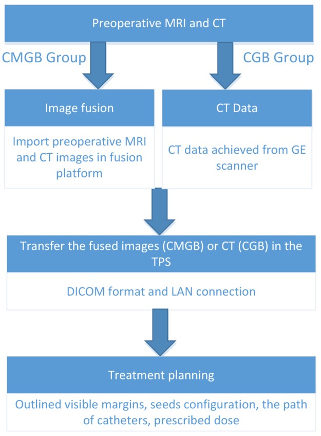
To develop a fast, accurate and robust method of fusing Computed Tomography (CT) with pre-operative Magnetic Resonance Imaging (MRI) and evaluate the impact of using the fused data on the implantation of Iodine-125 (I) seeds for brachytherapy of high-grade gliomas (HGG).
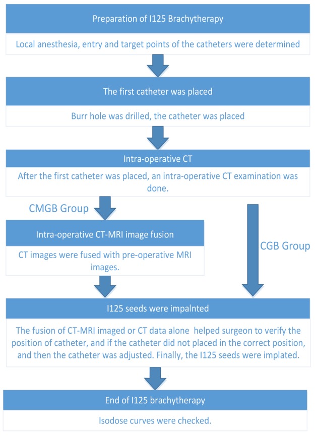
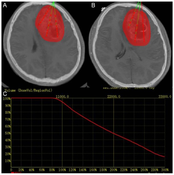
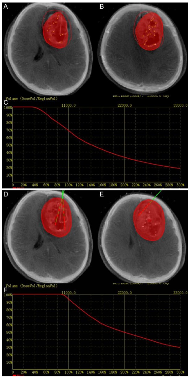
A study was performed on a cohort of 10 consecutive patients with HGG were treated by I brachytherapy with CT-MRI fusion image guided (CMGB), and 10 patients treated with CT alone guided (CGB). Statistical analysis was performed to compare (1) the planning target volume, (2) the accuracy of location of catheters, (3) the target volume covered by 150% prescribe dose (), (4) the target volume covered by 200% prescribe dose (), and (5) the conformity index () with or without fused data.
The median planning target volume was 50.1 cm in CGB, and 56.25 cm in CMGB with significant difference ( = 0.005). The accuracy of catheter insertion was 94.4% with CMGB and 78.9% with CGB. The median and was 45.32% vs 64.24% and 32.81% vs 53.17% in CGB and CMGB, respectively. There was significant difference for (83.5% 74.5%, < 0.05) in the two groups for the post-operative verification.
The proposed MRI-CT fusion method enables a quantitative assessment of impact on HGG brachytherapy. The additional information obtained from the fused images can be utilized for more accurate delineation of lesion boundaries and targeting of catheters. Experimental results show that the fusion algorithm is robust and reliable in clinical practice.
开发一种快速、准确且稳健的将计算机断层扫描(CT)与术前磁共振成像(MRI)融合的方法,并评估使用融合数据对碘 - 125(I)种子植入用于高级别胶质瘤(HGG)近距离放射治疗的影响。
对连续10例接受I近距离放射治疗且采用CT - MRI融合图像引导(CMGB)的HGG患者以及10例仅采用CT引导(CGB)的患者进行了一项研究。进行统计分析以比较(1)计划靶体积,(2)导管定位的准确性,(3)150%处方剂量覆盖的靶体积(),(4)200%处方剂量覆盖的靶体积(),以及(5)有无融合数据时的适形指数()。
CGB组的中位计划靶体积为50.1 cm³,CMGB组为56.25 cm³,差异有统计学意义( = 0.005)。CMGB组导管插入的准确率为94.4%,CGB组为78.9%。CGB组和CMGB组的中位和分别为45.32%对64.24%以及32.81%对53.17%。术后验证两组的差异有统计学意义(83.5% 74.5%, < 0.05)。
所提出的MRI - CT融合方法能够对HGG近距离放射治疗的影响进行定量评估。从融合图像获得的额外信息可用于更准确地描绘病变边界和导管靶向。实验结果表明该融合算法在临床实践中稳健且可靠。