• 文献检索
  • 文档翻译
  • 深度研究
  • 学术资讯
  • Suppr Zotero 插件Zotero 插件
  • 邀请有礼
  • 套餐&价格
  • 历史记录
应用&插件
Suppr Zotero 插件Zotero 插件浏览器插件Mac 客户端Windows 客户端微信小程序
定价
高级版会员购买积分包购买API积分包
服务
文献检索文档翻译深度研究API 文档MCP 服务
关于我们
关于 Suppr公司介绍联系我们用户协议隐私条款
关注我们

Suppr 超能文献

核心技术专利:CN118964589B侵权必究
粤ICP备2023148730 号-1Suppr @ 2026

文献检索

告别复杂PubMed语法,用中文像聊天一样搜索,搜遍4000万医学文献。AI智能推荐,让科研检索更轻松。

立即免费搜索

文件翻译

保留排版,准确专业,支持PDF/Word/PPT等文件格式,支持 12+语言互译。

免费翻译文档

深度研究

AI帮你快速写综述,25分钟生成高质量综述,智能提取关键信息,辅助科研写作。

立即免费体验

垂体黄色肉芽肿:临床特征、影像学表现和术后转归。

Pituitary xanthogranulomas: clinical features, radiological appearances and post-operative outcomes.

机构信息

B4 Office, Department of Neurosurgery, University Hospital of Wales, Cardiff, CF 14 4XW, UK.

Department of Endocrinology, University Hospital of Wales, Cardiff, CF 14 4XW, UK.

出版信息

Pituitary. 2018 Jun;21(3):256-265. doi: 10.1007/s11102-017-0859-x.

DOI:10.1007/s11102-017-0859-x
PMID:29363000
原文链接:https://pmc.ncbi.nlm.nih.gov/articles/PMC5942345/
Abstract

BACKGROUND

Xanthogranulomas are inflammatory masses most commonly found at peripheral sites such as the skin. Sellar and parasellar xanthogranulomas are rare and present a diagnostic challenge as they are difficult to differentiate from other sellar lesions such as craniopharyngiomas and Rathke's cleft cysts pre-operatively. Their radiological imaging features are yet to be clearly defined, and clinical outcomes after surgery are also uncertain. This study reviews clinical presentation, radiological appearances, and clinical outcomes in a cohort of patients with pituitary xanthogranulomas.

METHODS

A prospectively maintained pituitary surgery database was screened for histologically confirmed pituitary xanthogranulomas between May 2011-December 2016. Retrospective case note assessments were then performed by three independent reviewers. Patient demographics, clinical presentations, imaging, and clinical outcomes were analysed.

RESULTS

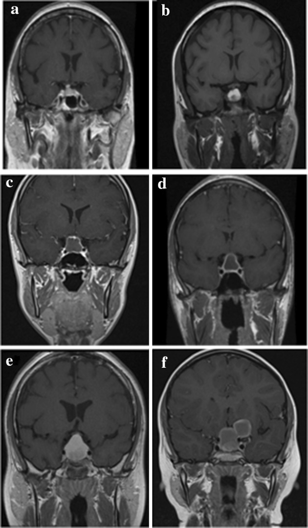
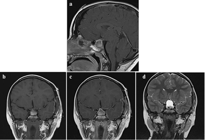
During the study period 295 endoscopic endonasal pituitary surgeries were performed. Six patients had confirmed pituitary xanthogranulomas (2%). Patients most commonly presented with visual field deficits and/or endocrine dysfunction. Common imaging features included: a cystic consistency, hyperintensity on T1-weighted MR images, and contrast enhancement either peripherally (n = 3) or homogenously (n = 3). The most common pre-operative endocrine deficits were hyperprolactinaemia and hypoadrenalism (at least one of which was identified in 4/6 patients; 66%). Thirty-three percent (2/6) of patients presented with diabetes insipidus. The most common post-operative endocrinological deficits were adrenocortical dysfunction (66%) and gonadotropin deficiency (66%). Visual assessments normalised in all six patients post-operatively. Gross total resection was achieved in all patients, and at median follow up of 33.5 months there were no cases of tumour recurrence.

CONCLUSIONS

The prevalence of pituitary xanthogranulomas in our series is higher than that suggested in the literature. Surgery restored normal vision to all cases, however four patients (67%) required long-term hormonal replacement post-operatively. Imaging features such peripheral rim enhancement, a suprasellar tumour epicentre, and the absence of both calcification or cavernous sinus invasion were identified as potential indicators that together should alert clinicians to the possibility of pituitary xanthogranuloma when assessing patients with cystic sellar and parasellar tumours.

摘要

背景

黄色肉芽肿是最常见于皮肤等外周部位的炎症性肿块。鞍上和鞍旁黄色肉芽肿罕见,术前难以与颅咽管瘤和 Rathke 裂囊肿等其他鞍内病变区分,诊断具有挑战性。其影像学表现尚未明确,术后临床结果也不确定。本研究回顾了一组垂体黄色肉芽肿患者的临床表现、影像学表现和临床结果。

方法

2011 年 5 月至 2016 年 12 月,通过前瞻性维持的垂体手术数据库筛选出经组织学证实的垂体黄色肉芽肿患者。然后由三名独立审查员对病历进行回顾性评估。分析患者的人口统计学资料、临床表现、影像学表现和临床结果。

结果

研究期间共进行了 295 例内镜经鼻垂体手术。6 例患者被证实患有垂体黄色肉芽肿(2%)。患者最常见的表现为视野缺损和/或内分泌功能障碍。常见的影像学特征包括:囊性一致性、T1 加权磁共振成像上的高信号强度,以及外周(n=3)或均匀(n=3)强化。最常见的术前内分泌缺陷是高泌乳素血症和肾上腺功能减退(至少有一项在 6/6 例患者中被识别;66%)。33%(2/6)的患者出现尿崩症。最常见的术后内分泌缺陷是肾上腺皮质功能障碍(66%)和促性腺激素缺乏(66%)。所有患者术后视力均恢复正常。所有患者均达到大体全切除,中位随访 33.5 个月后无肿瘤复发病例。

结论

本研究中,垂体黄色肉芽肿的患病率高于文献报道。手术使所有病例的视力恢复正常,但 4 例患者(67%)术后需要长期激素替代治疗。外周边缘强化、鞍上肿瘤中心、无钙化或海绵窦侵犯等影像学特征被认为是潜在的指标,这些指标结合起来,在评估囊性鞍内和鞍旁肿瘤的患者时,应能提醒临床医生注意垂体黄色肉芽肿的可能性。

https://cdn.ncbi.nlm.nih.gov/pmc/blobs/c808/5942345/97c3656cac07/11102_2017_859_Fig3_HTML.jpg
https://cdn.ncbi.nlm.nih.gov/pmc/blobs/c808/5942345/eea78e806a66/11102_2017_859_Fig1_HTML.jpg
https://cdn.ncbi.nlm.nih.gov/pmc/blobs/c808/5942345/d5b922d49a67/11102_2017_859_Fig2_HTML.jpg
https://cdn.ncbi.nlm.nih.gov/pmc/blobs/c808/5942345/97c3656cac07/11102_2017_859_Fig3_HTML.jpg
https://cdn.ncbi.nlm.nih.gov/pmc/blobs/c808/5942345/eea78e806a66/11102_2017_859_Fig1_HTML.jpg
https://cdn.ncbi.nlm.nih.gov/pmc/blobs/c808/5942345/d5b922d49a67/11102_2017_859_Fig2_HTML.jpg
https://cdn.ncbi.nlm.nih.gov/pmc/blobs/c808/5942345/97c3656cac07/11102_2017_859_Fig3_HTML.jpg

相似文献

1
Pituitary xanthogranulomas: clinical features, radiological appearances and post-operative outcomes.垂体黄色肉芽肿:临床特征、影像学表现和术后转归。
Pituitary. 2018 Jun;21(3):256-265. doi: 10.1007/s11102-017-0859-x.
2
Parasellar xanthogranulomas.鞍旁黄色瘤
J Neurosurg. 2015 Apr;122(4):812-7. doi: 10.3171/2014.12.JNS14542. Epub 2015 Jan 23.
3
Clinicoradiologic Features and Surgical Outcomes of Sellar Xanthogranulomas: A Single-Center 10-Year Experience.鞍区黄色肉芽肿的临床放射学特征及手术结果:单中心10年经验
World Neurosurg. 2017 Mar;99:439-447. doi: 10.1016/j.wneu.2016.12.017. Epub 2016 Dec 18.
4
Cholesterol granulomas presenting as sellar masses: a similar, but clinically distinct entity from craniopharyngioma and Rathke's cleft cyst.表现为鞍区肿块的胆固醇肉芽肿:一种与颅咽管瘤和拉克氏囊肿相似但临床特征不同的实体。
Pituitary. 2017 Jun;20(3):325-332. doi: 10.1007/s11102-016-0775-5.
5
Rathke's cleft cysts: differentiation from other cystic lesions in the pituitary fossa by use of single-shot fast spin-echo diffusion-weighted MR imaging.拉克氏裂囊肿:通过单次激发快速自旋回波扩散加权磁共振成像与垂体窝其他囊性病变相鉴别
Acta Neurochir (Wien). 2007 Aug;149(8):759-69; discussion 769. doi: 10.1007/s00701-007-1234-x. Epub 2007 Jul 9.
6
Xanthogranuloma of the Sellar Region: A Comprehensive Review of Neuroimaging in a Rare Inflammatory Entity.鞍区黄色肉芽肿:一种罕见炎症性病变的神经影像学综合综述
J Pers Med. 2022 Jun 8;12(6):943. doi: 10.3390/jpm12060943.
7
Clinical, radiological and pathological features of patients with Rathke's cleft cysts: tumors that may recur.拉克氏裂囊肿患者的临床、放射学及病理学特征:可能复发的肿瘤
J Clin Endocrinol Metab. 1997 Jul;82(7):2357-62. doi: 10.1210/jcem.82.7.4043.
8
Magnetic resonance imaging after transsphenoidal surgery of clinically non-functional pituitary macroadenomas and its impact on detecting residual adenoma.临床无功能垂体大腺瘤经蝶窦手术后的磁共振成像及其对检测残留腺瘤的影响。
Acta Neurochir (Wien). 2002 May;144(5):433-43. doi: 10.1007/s007010200064.
9
Diabetes insipidus secondary to sellar/parasellar lesions.继发于鞍区/鞍旁病变的尿崩症。
J Neuroendocrinol. 2021 Mar;33(3):e12954. doi: 10.1111/jne.12954.
10
Pituitary magnetic resonance imaging for sellar and parasellar masses: ten-year experience in 2598 patients.鞍区和鞍旁肿块的垂体磁共振成像:2598 例患者 10 年经验。
J Clin Endocrinol Metab. 2011 Jun;96(6):1633-41. doi: 10.1210/jc.2011-0168. Epub 2011 Apr 6.

引用本文的文献

1
Sellar Xanthogranulomatosis in a Two-Year-Old Girl: A Case Report.一名两岁女童的鞍区黄色肉芽肿:病例报告
Cureus. 2023 Nov 25;15(11):e49405. doi: 10.7759/cureus.49405. eCollection 2023 Nov.
2
Sellar xanthogranuloma as a diagnostic challenge: a report on five cases.鞍区黄色肉芽肿——一项诊断挑战:5例报告
Front Neurosci. 2023 Sep 22;17:1227144. doi: 10.3389/fnins.2023.1227144. eCollection 2023.
3
Inflammatory and Infectious Disorders in Endocrine Pathology.内分泌病理学中的炎症和感染性疾病。

本文引用的文献

1
Review of xanthomatous lesions of the sella.蝶鞍部黄色瘤样病变的综述。
Brain Pathol. 2017 May;27(3):377-395. doi: 10.1111/bpa.12498.
2
Cholesterol granulomas presenting as sellar masses: a similar, but clinically distinct entity from craniopharyngioma and Rathke's cleft cyst.表现为鞍区肿块的胆固醇肉芽肿:一种与颅咽管瘤和拉克氏囊肿相似但临床特征不同的实体。
Pituitary. 2017 Jun;20(3):325-332. doi: 10.1007/s11102-016-0775-5.
3
The sellar and suprasellar region: A "hideaway" of rare lesions. Clinical aspects, imaging findings, surgical outcome and comparative analysis.
Endocr Pathol. 2023 Dec;34(4):406-436. doi: 10.1007/s12022-023-09771-3. Epub 2023 May 20.
4
Pituitary adenoma associated with xanthogranuloma.垂体腺瘤伴黄色肉芽肿
Radiol Case Rep. 2023 Apr 5;18(6):2078-2080. doi: 10.1016/j.radcr.2023.03.015. eCollection 2023 Jun.
5
Intrasellar xanthogranuloma mimicking macroadenoma.鞍内黄色肉芽肿,类似于大腺瘤。
Neuroradiol J. 2024 Feb;37(1):123-125. doi: 10.1177/19714009231166075. Epub 2023 Mar 23.
6
Xanthogranuloma of the sellar region: a systematic review.鞍区黄色肉芽肿:系统评价。
Hormones (Athens). 2023 Jun;22(2):199-210. doi: 10.1007/s42000-023-00432-y. Epub 2023 Jan 25.
7
Successful immunomodulatory treatment for recurrent xanthogranulomatous hypophysitis in an adolescent: illustrative case.青少年复发性黄色肉芽肿性垂体炎的成功免疫调节治疗:病例说明
J Neurosurg Case Lessons. 2022 Aug 29;4(9):CASE22191. doi: 10.3171/CASE22191.
8
Xanthogranuloma of the Sellar Region: A Comprehensive Review of Neuroimaging in a Rare Inflammatory Entity.鞍区黄色肉芽肿:一种罕见炎症性病变的神经影像学综合综述
J Pers Med. 2022 Jun 8;12(6):943. doi: 10.3390/jpm12060943.
9
Sellar xanthogranuloma: A diagnostic challenge.鞍区黄色肉芽肿:一项诊断挑战。
Surg Neurol Int. 2022 Mar 4;13:76. doi: 10.25259/SNI_1180_2021. eCollection 2022.
10
Xanthogranuloma of the sellar region: A case report.鞍区黄色肉芽肿:一例报告。
Medicine (Baltimore). 2020 Oct 2;99(40):e22619. doi: 10.1097/MD.0000000000022619.
鞍区和鞍上区:罕见病变的“隐匿之处”。临床情况、影像学表现、手术结果及对比分析。
Clin Neurol Neurosurg. 2016 Oct;149:154-65. doi: 10.1016/j.clineuro.2016.08.011. Epub 2016 Aug 12.
4
Calcified Suprasellar Xanthogranuloma Presenting with Primary Amenorrhea in a 17-Year-Old Girl: Case Report and Literature Review.钙化性鞍上黄色瘤在一名17岁女孩中表现为原发性闭经:病例报告及文献综述
World Neurosurg. 2015 Sep;84(3):866.e11-4. doi: 10.1016/j.wneu.2015.04.024. Epub 2015 Apr 25.
5
Parasellar xanthogranulomas.鞍旁黄色瘤
J Neurosurg. 2015 Apr;122(4):812-7. doi: 10.3171/2014.12.JNS14542. Epub 2015 Jan 23.
6
Clinicopathological features of sellar region xanthogranuloma: correlation with Rathke's cleft cyst.鞍区黄色肉芽肿的临床病理特征:与 Rathke 裂隙囊肿的相关性。
Brain Tumor Pathol. 2013 Oct;30(4):233-41. doi: 10.1007/s10014-012-0130-0. Epub 2013 Jan 16.
7
Sterile brain abscess due to juvenile xanthogranuloma: DWI characteristics.青少年黄色肉芽肿所致无菌性脑脓肿:扩散加权成像特征
J Neuroimaging. 2013 Jul;23(3):437-40. doi: 10.1111/j.1552-6569.2012.00725.x. Epub 2012 Jul 12.
8
Xanthogranuloma of the intrasellar region presenting in pituitary dysfunction: a case report.以垂体功能障碍为表现的鞍内区黄色肉芽肿:一例报告
J Med Case Rep. 2012 Apr 29;6:119. doi: 10.1186/1752-1947-6-119.
9
Xanthogranuloma of the sellar region in a patient with sarcoidosis.一名结节病患者的鞍区黄色肉芽肿。
Acta Clin Croat. 2010 Mar;49(1):61-5.
10
Endocrinological and MRI features of pituitary adenomas with marked xanthogranulomatous reaction.伴有明显黄色肉芽肿反应的垂体腺瘤的内分泌和 MRI 特征。
Neuroradiology. 2010 Nov;52(11):997-1002. doi: 10.1007/s00234-010-0675-8. Epub 2010 Mar 18.