Villemain Olivier, Robin Justine, Bel Alain, Kwiecinski Wojciech, Bruneval Patrick, Arnal Bastien, Rémond Mathieu, Tanter Mickael, Messas Emmanuel, Pernot Mathieu
Institut Langevin, ESPCI, CNRS, Inserm U979, PSL Research University, Paris, France.
Hôpital Européen Georges Pompidou, Université Paris Descartes, Cardio-Vascular Departement, UMR 970, Paris, France.
JACC Basic Transl Sci. 2017 Aug;2(4):372-383. doi: 10.1016/j.jacbts.2017.03.012.
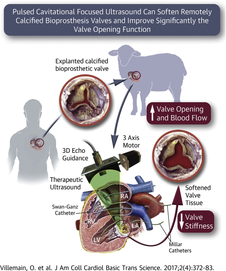
The majority of prosthetic heart valves currently implanted are tissue valves that can be expected to calcify with time and eventually fail. Surgical or percutaneous redux valve replacement is associated with higher rate of complications. We propose a novel non-invasive therapeutic approach based on the use of pulsed cavitational ultrasound (PCU) to improve the valvular function of degenerative calcified bioprosthesis.
Our study aims to demonstrate in vitro and in vivo on an ovine model that PCU can significantly improve the bioprosthesis opening by softening remotely the calcified stiff cusps.
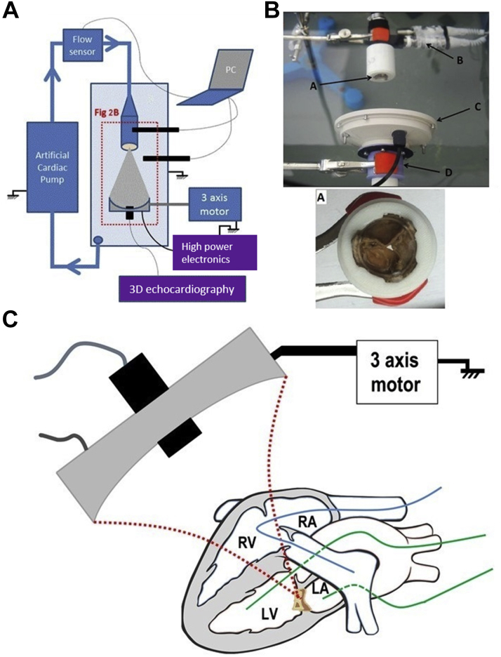
All the experiments were performed on calcified bioprosthetic valves explanted from human patients. PCU was performed in vitro on calcified bioprosthesis mounted on a hydraulic bench with pulsatile flow (n=8) and in vivo on an ovine model with implanted calcified bioprosthesis (n=7). We used 3D echocardiography, pressure and flow sensors, quantitative stiffness evaluation using shear wave elastography, micro-CT imaging and histology to evaluate in vitro and in vivo the effect of PCU.
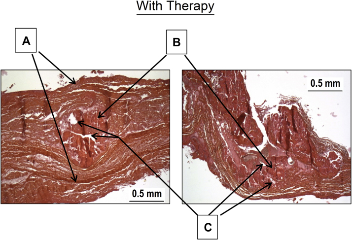
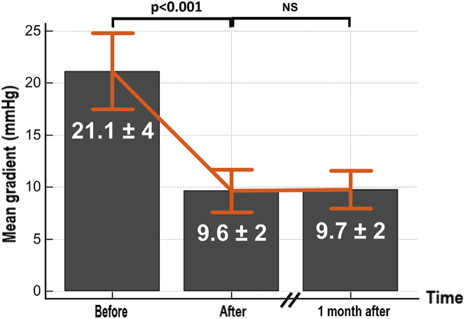
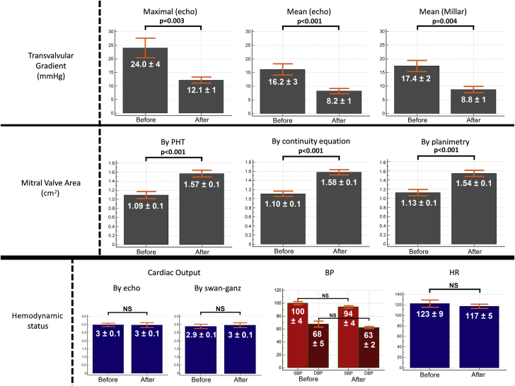
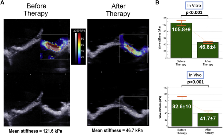
The transvalvular gradient was found to decrease by a mean of 50% after PCU in both in vitro (from 21.1±3.9 to 9.6±1.7 mmHg, p<0.001) and in vivo setup (from 16.2±3.2 to 8.2±1.3 mmHg, p<0.001), with a decrease of valve stiffness (in vitro: from 105.8±9 to 46.6±4 kPa, p<0.001; in vivo: from 82.6±10 to 41.7±7 kPa, p<0.001) and an increase of valve area (from 1.10±0.1 to 1.58±0.1 cm p<0.001). Histology and micro-CT imaging showed modifications of calcification structure without loss of calcification volume or alteration of the leaflet superficial structures.
We have demonstrated in vitro and in vivo that PCU can decrease a calcified bioprosthesis stenosis by softening the leaflets remotely. This new non-invasive approach has the potential to improve the outcome of patients with severe bioprosthesis stenosis.
目前植入的大多数人工心脏瓣膜是组织瓣膜,随着时间的推移可能会钙化并最终失效。手术或经皮再次瓣膜置换术的并发症发生率较高。我们提出了一种基于脉冲空化超声(PCU)的新型非侵入性治疗方法,以改善退行性钙化生物假体的瓣膜功能。
我们的研究旨在在体外和绵羊模型体内证明,PCU可通过远程软化钙化僵硬的瓣叶显著改善生物假体的开放。
所有实验均在从人类患者身上取出的钙化生物假体瓣膜上进行。PCU在体外对安装在具有脉动流的液压工作台上的钙化生物假体进行(n = 8),在体内对植入钙化生物假体的绵羊模型进行(n = 7)。我们使用三维超声心动图、压力和流量传感器、使用剪切波弹性成像进行定量硬度评估、微型计算机断层扫描成像和组织学来评估PCU在体外和体内的效果。
在体外(从21.1±3.9降至9.6±1.7 mmHg,p<0.001)和体内设置(从16.2±3.2降至8.2±1.3 mmHg,p<0.001)中,PCU后跨瓣膜梯度平均降低50%,瓣膜硬度降低(体外:从105.8±9降至46.6±4 kPa,p<0.001;体内:从82.6±10降至41.7±7 kPa,p<0.001),瓣膜面积增加(从1.10±0.1增至1.58±0.1 cm,p<0.001)。组织学和微型计算机断层扫描成像显示钙化结构有改变,但钙化体积未减少,瓣叶表面结构未改变。
我们已在体外和体内证明,PCU可通过远程软化瓣叶降低钙化生物假体狭窄程度。这种新的非侵入性方法有可能改善严重生物假体狭窄患者的治疗效果。